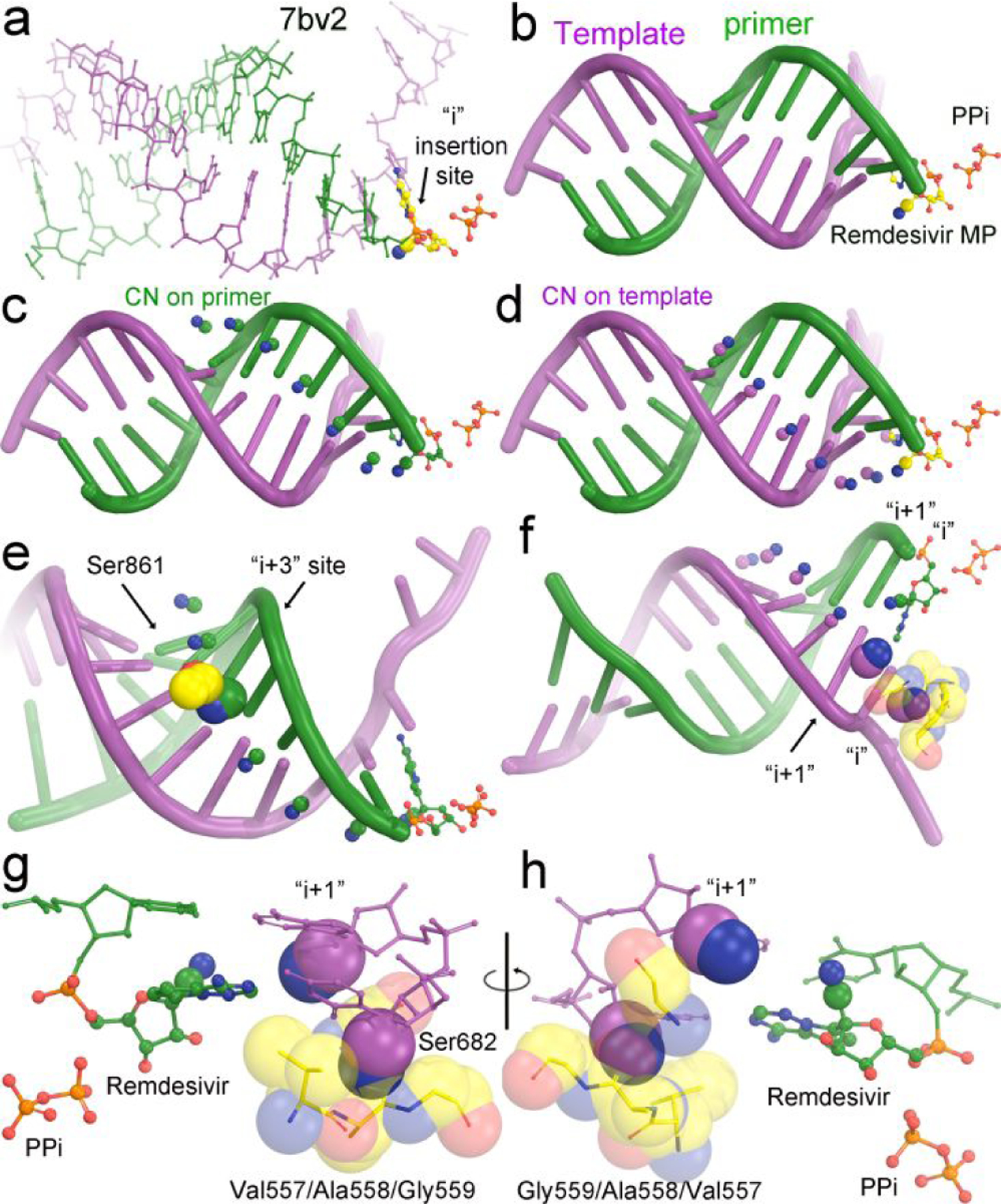
Figure 2.

Modeling of the cyano substitutions at various positions of primer and template nucleotides. (a, b) Experimental structure of the 7bv2 complex with pyrophosphate and RMP in colored ball-and-stick model. The cyano group is shown in 30% of van der Waals radii. (c) Modeling of the cyano substitutions on the nucleotides of the primer strand based on (i) tetrahedral geometry of C1’ atom, (ii) the C-C single bond of 1.47 Å, and (iii) the C-N triple bond of 1.14 Å. (d) Modeling of the cyano substitutions on the nucleotides of the template strand. (e) Known clashes between the sidechain of Ser861 of RdRp and the cyano substitution during the translocation from the i+3 site to the i+4 site. (f) New severe clashes between the cyano substitution of the template nucleotide at the i site and backbone carbonyl group of Val557 of RdRp and a minor clash between the cyano substitution of the template nucleotide at the i+1 site and the backbone carbonyl of Ser682 of RdRp. (g,h) Close-up views of the first two base pairs in 180° orientation of (f).
