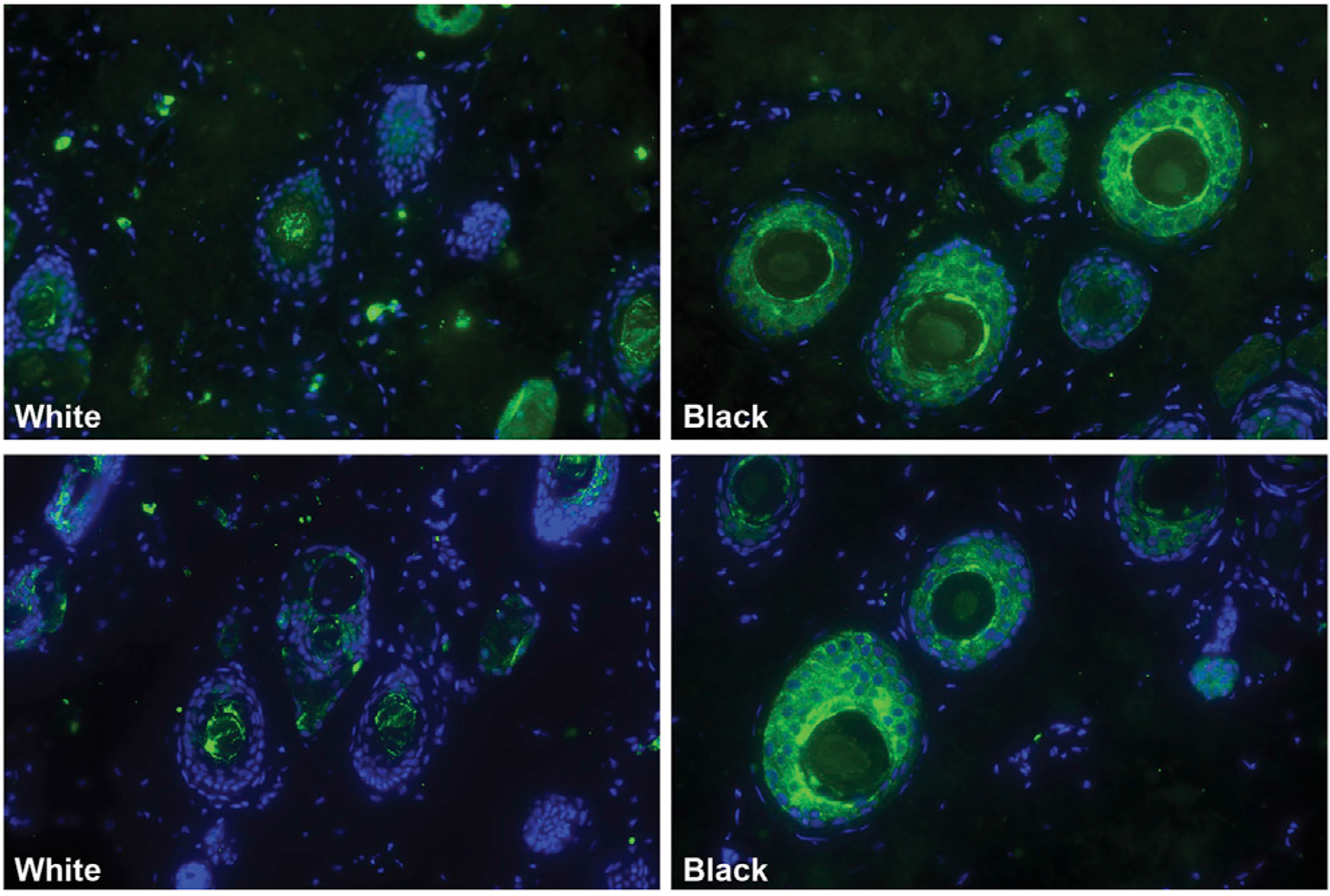
Figure 2. The White Facial Patch Contains a Paucity of Melanocytes Relative to the Adjacent Dark Fur.

This supports the neural crest cell hypothesis for domestication phenotypes. Green indicates the presence of melanocytes (Trp1 antibody); the blue is a nuclear stain. Samples from two different marmosets are depicted, one per row.
