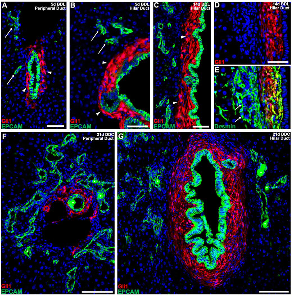
Figure 4. Gli1+ PMCs do not wrap around epithelial cells of the ductular reaction.
(A, B) 5 days after BDL, Gli1+ PMCs continue to expand around cholangiocytes of main peripheral (A) and hilar (B) ducts (arrowheads) with EPCAM+ cells of the ductular reaction forming without any communicating Gli1+ PMCs (arrows). (C) 14 days after BDL, a clear distinction between Gli1+ PMCs of the bile duct (arrowheads) and EPCAM+ cells of the ductular reaction is still detected. (D, E) Cells of the ductular reaction are surrounded by desmin+ stellate cells. (F, G) After 21 days of DDC, Gli1+ PMCs consistently surround the main peripheral (F) and hilar (G) ducts, without surrounding E PCAM+ cells of the ductular reaction. (n = 4 for all experiments). * denotes autofluorescent porphyrin pigment plugs. Scale bars, 50 μm (A-E); 100 μm (F, G).

