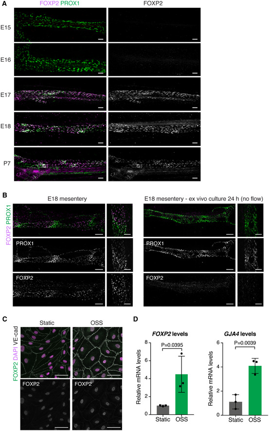
Figure 2. FOXP2 expression is regulated by flow.

-
AWhole‐mount immunofluorescence of embryonic mesenteries of the indicated developmental stages. Note induction of FOXP2 expression at E17.
-
BWhole‐mount immunofluorescence of E18 mesenteries fixed immediately after dissection (left panel) or after 24 h of ex vivo culture (right panel). Note loss of patterning of PROX1high valve LECs and downregulation of FOXP2 expression in flow‐abrogated vessels after ex vivo culture.
-
C, DImmunofluorescence (C) and qRT–PCR analysis (D) of HDLECs grown under static conditions or exposed to OSS for 48 h (n = 3 independent experiments). Data are presented as mean ± SD. P, Student's t‐test.
Data information: Scale bar: 100 µm (A, B), 50 μm (C).
Source data are available online for this figure.
