
Figure 6.
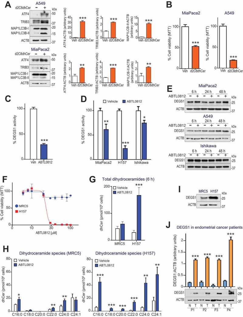
Dihydroceramide accumulation by ABTL0812 treatment induces ER stress, autophagy and selective cancer cell death. (a) Cells were treated with 25 µmol/L of the dideuterated dihydroceramide analog d2C8dhCer for 48 h, then lysed. The levels of ER stress markers ATF4 and TRIB3, as well as MAP1LC3B lipidation, were monitored by immunoblotting. Histograms show quantification of ATF4, TRIB3 and MAP1LC3B-II levels normalized to ACTB levels and represent the mean fold-change relative to vehicle-treated cells. (b) MiaPaca2 and A549 cells were treated 48 h with 25 µmol/L of d2c8dhCer, and cell viability was determined by MTT assay. Each value is the mean ± SD of two different experiments performed in triplicates. (c,d) ABTL0812 treatment induces DEGS1 inhibition. (c) A549 cell lysates were treated with vehicle or 100 µmol/L ABTL0812 for 4 h. (d) Cultured MiaPaca2 (pancreatic), H157 (SNSCLC) and Ishikawa (endometrial) cancer cells were treated 6 h with vehicle or 100 µmol/L ABTL0812. DEGS1 activity was determined as described in the Materials and Methods Section using DHCerC6NBD as substrate. Data expressed in percentage of the CerC6NBD pick area. Each value is the mean ± SD of three different determinations. (e) DEGS1 levels visualized by immunoblotting. (f) Selectivity of ABTL0812 cytotoxic effect for cancer cells. Human lung fibroblast MRC5 and squamous NSCLC H157 cells were treated with ABTL0812 for 48 h, and cell viability determined by MTT assay. Results representative of three separate experiments. (g,h) ABTL0812 does not induce accumulation of dihydroceramides in a non-tumoral cell line. (g) Levels of total dihydroceramides in MRC5 fibroblasts and H157 cancer cells, after 6 h of ABTL0812 treatment. (h) Levels of molecular dihydroceramide species. Each value is the mean ± SD of three different determinations. (i) DEGS1 protein levels (immunoblot) in MRC5 and H157 cells. (j) Human endometrial tumors express higher levels of DEGS1 protein than adjacent non-tumoral tissue. Immunoblot analysis of samples (tumor area [T] and non-tumor area [N]) from 4 different patients with endometrial cancer. *, P < 0.05; **, P < 0.005; ***, P < 0.001 from vehicle-treated cells