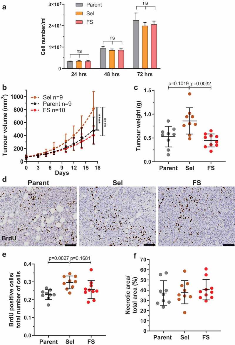
Figure 6.

Pore-selected MDA MB 231 breast cancer cells grow faster as orthotopic xenograft tumours. (a). Proliferation of Parent, Sel and FS MDA 231 cells was determined by plating 30,000 cells per well of 12-well plate and counting cell numbers after 24, 48 and 72 hours. The number of experimental replicates were n = 12 for each population at each time point. Kruskal-Wallis test with Dunn’s multiple comparisons. ns = not significant. Means ± SEM. (b). Parent, Sel and FS cells were injected at 106 cells in 50 µl PBS:matrigel into the fourth abdominal fat pad of female immuncompromised CD1-nu/nu mice. Tumour measurements from when they became palpable (day 0). Tumour volumes were quantified from calliper measurements using the formula 1/2(Length × Width2). Statistical analysis was by two-way ANOVA and post-hoc Tukey’s multiple comparison test. (**** = p < 0.0001). (c). Mean tumour weights (± SD) from each mouse after 17 days. Kruskal-Wallis test with Dunn’s multiple comparisons showing adjusted p values. (d). Representative images of anti-BrdU-stained tumour sections for Parent, Sel2 and FS1 MDA MB 231 tumours from mice that had been injected intraperitoneally with BrdU two hours prior to sacrifice. Scale bar = 100 µm. (e). Mean proportion of BrdU positive cells per total cells counted (± SD) in Parent, Sel2 and FS1 MDA MB 231 tumours. Kruskal-Wallis test with Dunn’s multiple comparisons showing adjusted p values. (f). Mean percentage necrotic area (± SD) determined visually in haematoxylin and eosin stained sections of Parent, Sel2 and FS1 MDA MB 231 tumours. Kruskal-Wallis test revealed no significant differences
