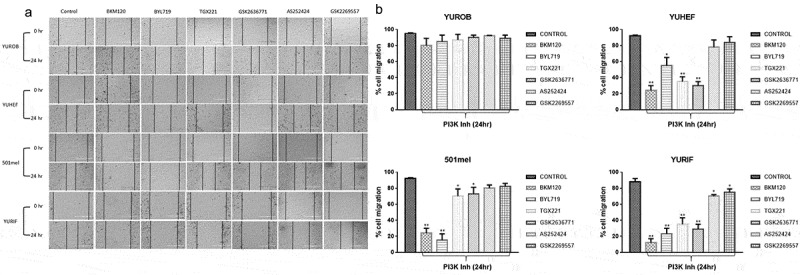
Figure 3.

Consequences of PI3K inhibitors on cell migration. A cell culture wound-healing assay was performed in six-well confluent plates. After creating a gap by scratching a confluent plate of cells, WT (YUROB), RAC1 (YUHEF), BRAF (501 mel) and BRAF/RAC1 (YURIF) cell lines were treated with 100 nM pan-PI3K (BKM120), PI3Kα (BYL719), PI3Kβ (TGX221, GSK2636771), PI3Kγ (AS252424) and PI3Kδ (GSK2269557) inhibitors. (a) Images were taken in an inverted microscope at 0 and 24 h. Scale bar 400 μm for all images. (b) Migration was quantified by measuring the length of the cell-free area. Maximum (100%) migration was determined by the control. Percentage of migration of each treated cell line was measured against untreated cells (control). The results are shown as mean (SD) of at least three independent experiments (* p < 0.05, ** p < 0.01 versus control)
