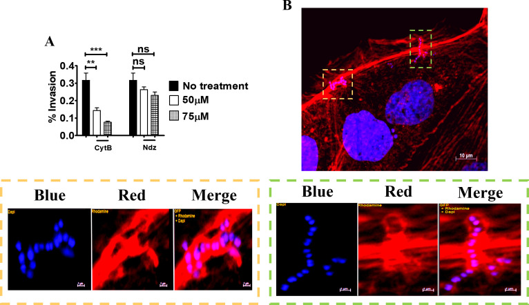
Fig 5. Actin-dependent invasion of Caco-2 cells by GBS.
(A) Effect of cytochalasin B (CytB) and nocodazole (Ndz) on GBS invasion of 5-day-old Caco-2 islets at the non-cytotoxic concentrations of 50 and 75μM, respectively. Shown are means ± SD of three independent experiments conducted in triplicate **, p<0.01; ***, p<0,001 by one-way Anova and Bonferroni test. (B) Fluorescence microscopy analysis of 5-day-old islets infected with GBS. Scale bar = 10μm. The bottom panels are magnifications of the areas indicated by the two coloured dashed rectangles (orange and green). Cell-associated streptococci appear to be enveloped by actin filaments (red) below the outer edges of the cells. Scale bar = 1μm. Nuclei and nucleoids were stained with DAPI (Blue), actin with Phalloidin-iFluor 555 (Red).

