
Figure 2.
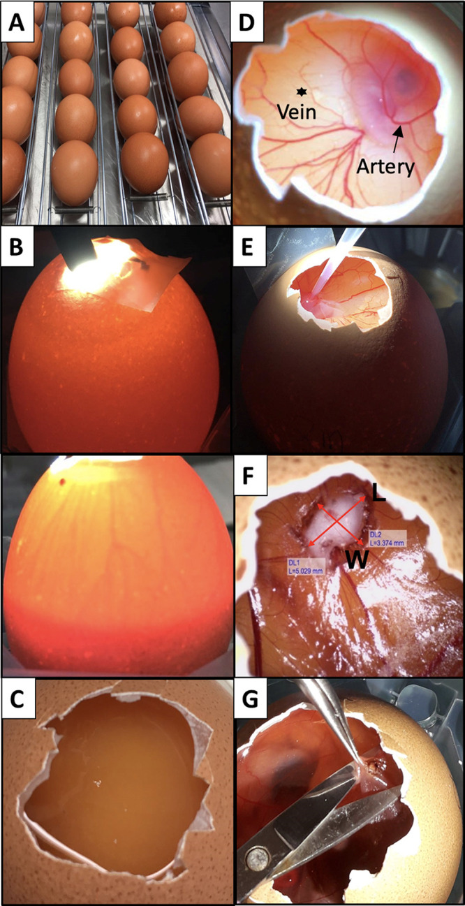
Images of the main steps of the CAM protocol. (A) Eggs are horizontally placed in the tilting trays before the incubation starts. (B) A light source placed on top/behind the egg allows identification of the location of the air chamber and the existence of a vascular network (Top, infertile egg; bottom, fertile egg). (C) An unfertilized egg is associated with the absence of the embryo and of the vascular network. (D) Distinction between artery (arrow) and vein (asterisk). Arteries are thicker and darker compared to veins.25 (E) Image showing the grafting procedure of SCC-25 cancer cells onto the bleeding blood vessel. (F) Tumor volume measurement determined by means of width and length. The length is associated with the longest diameter of the tumor mass. (G) Harvesting procedure of the tumor at EDD17. The CAM membrane is gently lifted with tweezers and the tumor is cut with scissors.