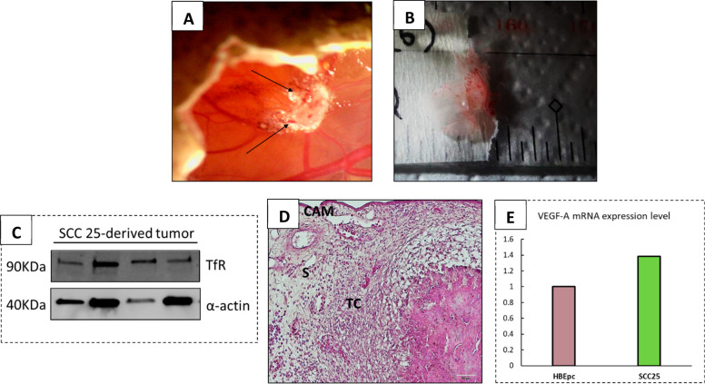
Figure 3.
Characterization of harvested SCC25 tumors. (A) Representative image of SCC-25 solid tumor grown onto the CAM. The arrows indicate the blood vessels across the tumor mass. (B) Example of solid and vascularized tumor harvested at EDD17. (C) Western blotting analysis depicting the expression of the TfR marker in SCC-25-tumor-derived cells. (D) H&E staining of SCC-25 tumor-derived cells showing the tissue structure and cells distribution (TC = tumor cells; S = stroma; scale bar = 20 μm). (E) Real-time PCR measurement of VEGF-A mRNA expression levels in SCC-25 cancer cells compared to HBEpc bronchial cell line.

