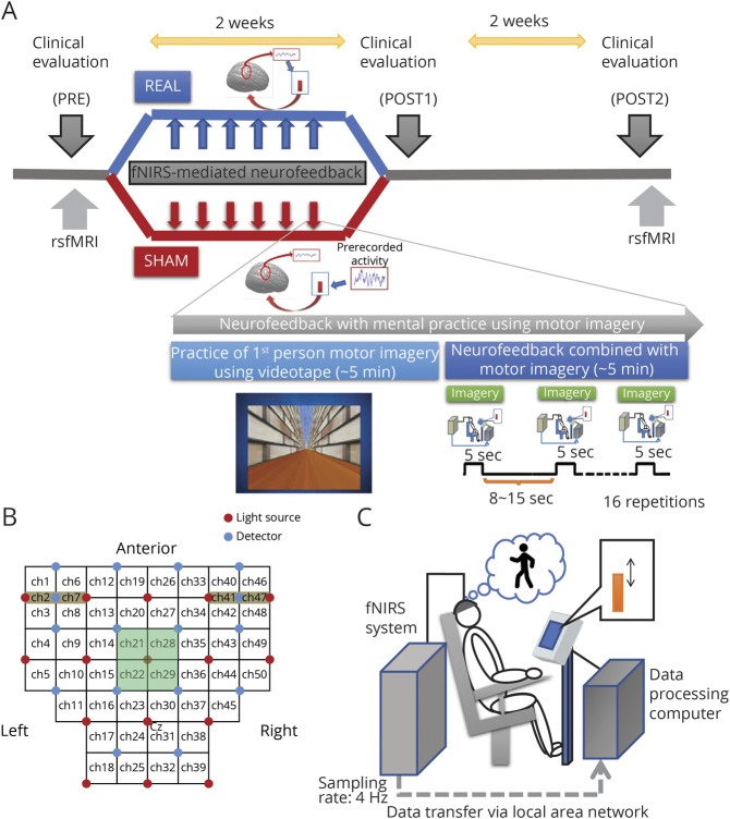
Figure 2. Study Protocol and Setting of the Functional Near Infrared Spectroscopy (fNIRS) Neurofeedback Session.
(A)Schematic illustration of the protocol for this study. Patients were randomly allocated to 2 groups, real feedback and sham feedback, and were subjected to 6 sessions of neurofeedback training combined with motor imagery. In the real group, the patients were provided with actual cortical activation during the motor imagery task, so they could learn how to regulate their cortical activation. In contrast, patients in the sham group were provided irrelevant information (other patients' recorded data) and therefore could not learn how to regulate their cortical activation. In the neurofeedback training sessions, patients were asked to practice the first-person motor imagery of the gait and balance task using videos. After practicing, the participants were requested to perform the motor imagery task without the video but with visual feedback of the vertical bar as a measure of appropriate supplementary motor area (SMA) activation. Neurofeedback practice was provided 3 times per week for 2 weeks, and clinical measures were evaluated before intervention (pre), just after intervention (post1), and 2 weeks postintervention (post2). (B) During neurofeedback sessions, participants' cortical activation was measured using fNIRS. fNIRS signals from the channels covering the fronto-parietal scalp and suspected to cover the SMA (channels 21, 22, 28, and 29) were used for real-time processing and calculation of the feedback signal. (C) Illustration depicts the experiment setup wherein a participant is sitting on a chair and performing a gait and balance–related motor imagery task. rsfMRI = resting-state functional MRI.

