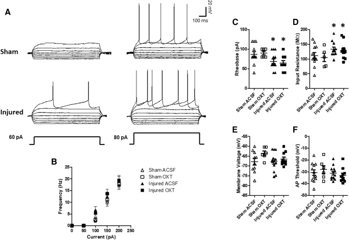
Figure 8.
OXT did not affect membrane properties of Layer II/III pyramidal neurons within the mPFC. Following behavioral testing, slices containing the mPFC were obtained at six to seven weeks after injury and neuronal activity was measured using whole-cell patch clamp electrophysiology as described in Materials and Methods. A, Representative current clamp traces from sham and brain-injured neurons. B, Frequency of neuron firing in response to varying levels of current injection. C, Rheobase. D, Input resistance. E, Membrane voltage. F, Spike threshold. Bars represent mean group values, and error bars represent SEM. Open triangles represent sham cells bathed with aCSF (N = 12), open squares represent sham cells bathed with 1 μm OXT (N = 6), filled triangles represent injured cells bathed with aCSF (N = 13), and filled squares represent injured cells bathed with OXT (N = 10); *p < 0.05.

