Figure 2.

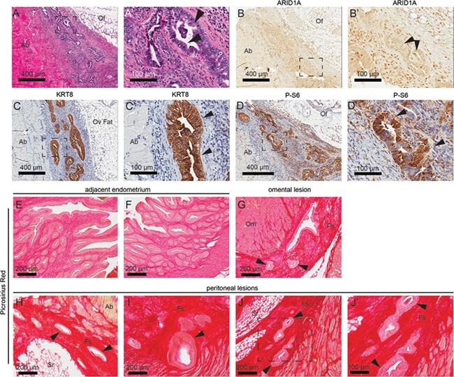
Endometrial glands formed in ovary are ARID1A-negative. (A) H&E staining in sections of ovarian lesion. (B–D) Immunohistochemistry (IHC) for ARID1A (B), KRT8 (ketatin 8) (C) and phospho-S6 (P-S6) (D) in sections of ovarian lesion. Panels A–D represent near-adjacent sections of extrauterine endometrial lesions, while panels A′–D′ represent portion of slide in panels A–D surrounded by black box. Arrows indicate epithelial gland formation. (E–J) Picrosirius red staining in sections of adjacent uterine tissue (E, F), omental lesions (G) and peritoneal lesions (H–J). Panel J′ represents portion in panel J surrounded by black box. Ab: abdominal wall; Cy: cyst; Fs: fibrotic stroma; Of: ovarian fat; Om: omentum; Oo: oocyte; Ov: ovary: Sr: serosa.
