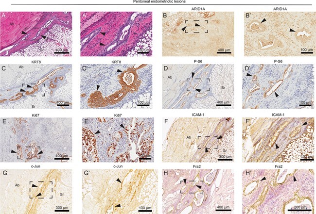
Figure 5.

Biomarker identification in peritoneal endometriotic lesions. (A) H&E staining of endometrial glands identified in the peritoneum. Endometriotic lesions were fused to the abdominal wall. (B–H) Immunohistochemical staining of peritoneal lesions for ARID1A (B), KRT8 (C), P-S6 (D), Ki67 (E), ICAM-1 (intercellular adhesion molecule 1) (F) and AP-1 (activator protein 1) subunits c-Jun (G) and Fra2 (Fos-related antigen 2) (H). Panels A–H represent near-adjacent sections of peritoneal endometriotic lesions, while panels A′–H′ represent portion of slide in panels A–H surrounded by black box. Arrows indicate endometrial glands. Ab: abdominal wall; Sr: serosa.
