Fig. 3.
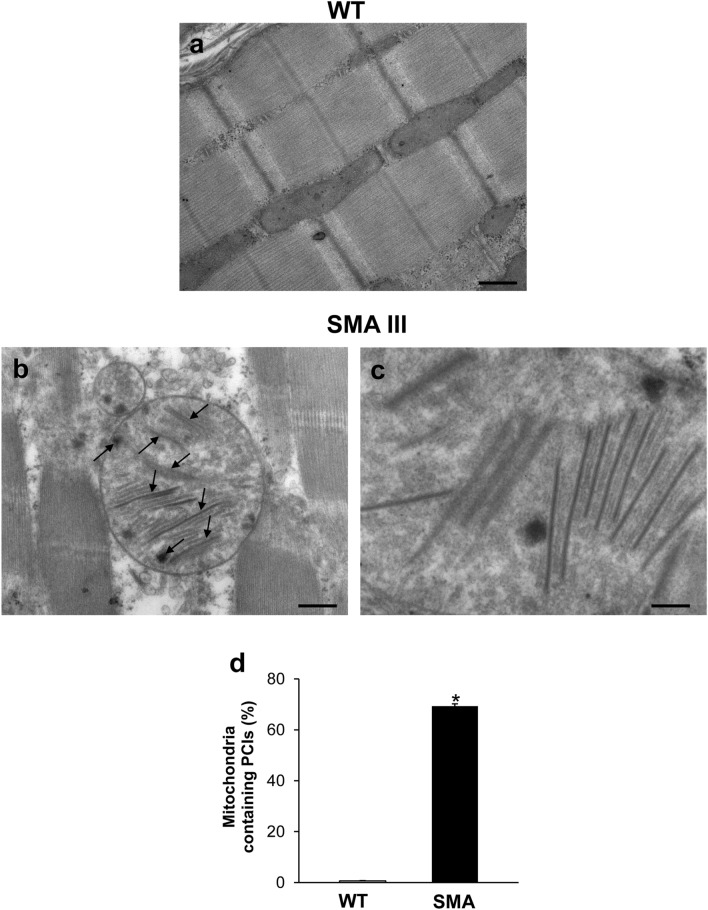
Representative TEM micrographs of mitochondrial paracrystalline inclusions (PCIs) occurring in the muscle of SMA mice. a, b Representative TEM micrographs showing mitochondrial paracrystalline inclusions (PCIs) in gastrocnemius muscle from WT and SMA mice. While PCIs are totally absent in the WT mouse (a), the mitochondria of the muscle from SMA mouse (b) were all filled with these rigid, rectangular, and electron-dense crystals (c). The graph d reports the percentage of mitochondria containing PCIs measured within the muscle of SMA compared with WT mice. Values are the mean percentage ± S.E.M. from 20 homogenous areas each measuring 6 μm2. Scale bar: a, b = 1 µm; c = 0.14 µm. *P ≤ 0.05 compared with WT

