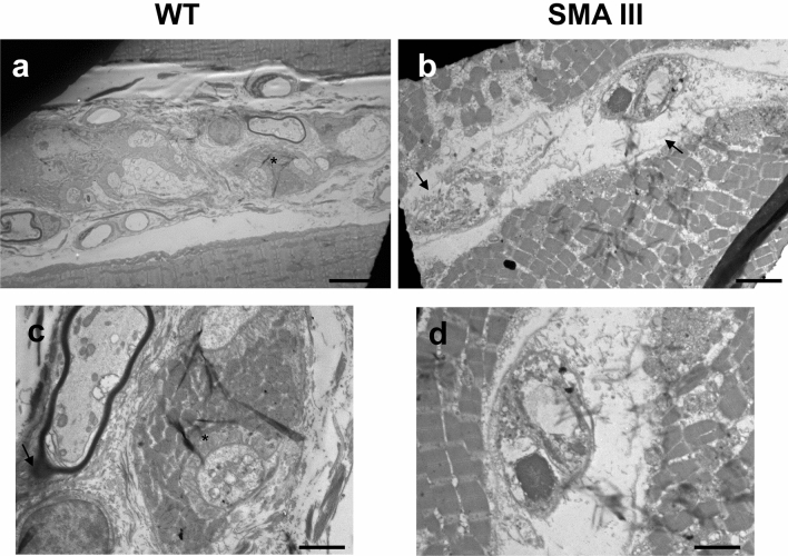
Fig. 4.
Representative TEM micrographs of muscle spindle from WT and SMA mice. a, c Representative TEM micrographs from WT mice showing muscle spindles with normal ultrastructure, concerning both intrafusal fibers with well-organized myofilaments, and regular capsule. At higher-magnification micrographs, one can appreciate well-conformed fibers receiving axon terminals (asterisk), which are surrounded by a well-organized layer of sarcoplasm. b, d Representative TEM micrographs from SMA mice showing dramatically altered muscle spindle architecture. Myofilaments are not recognizable, the capsule is thickened or disintegrated (arrow), and anomalous fibers with completely disarranged surrounding sarcoplasm appear. Scale bar: a, b = 18 µm; c, d = 3.5 µm

