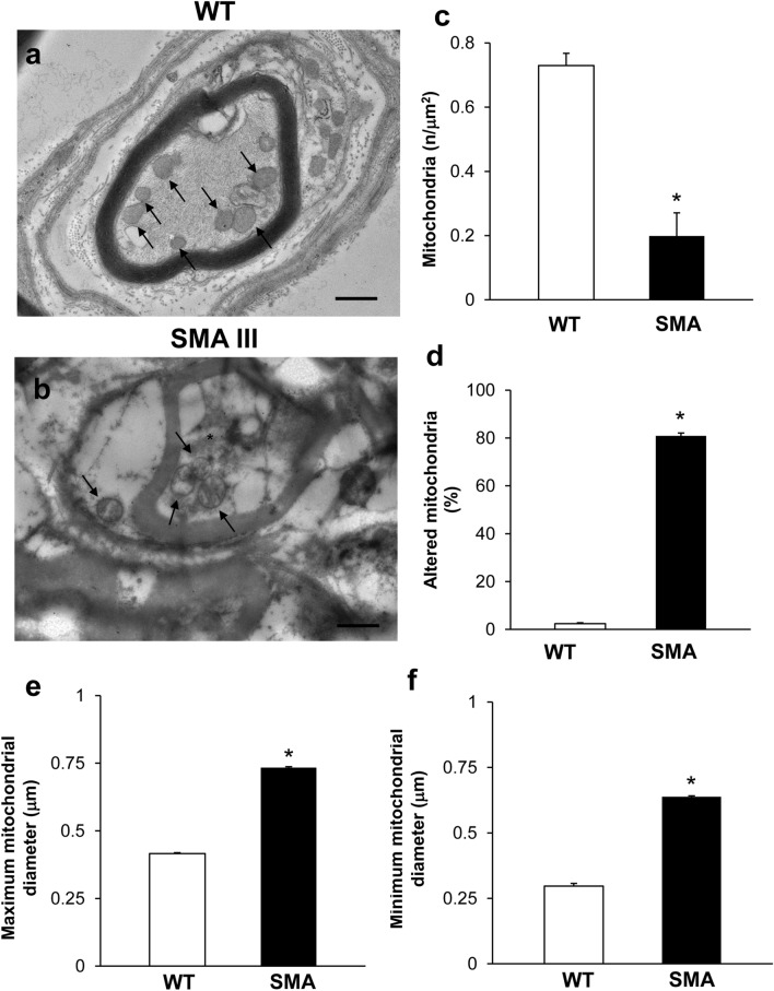
Fig. 5.
Representative electron micrographs of distal axons in WT and SMA mice. a Electron micrograph of cross-sections of myelinated axons from WT mice showing well-confirmed and organized myelinated sheath with axons lacking any obstructive materials within the axoplasm. A well-organized cytoplasm and healthy mitochondria are distinguishable in the axoplasm (arrows). b Electron micrograph of cross-sections of myelinated axons from SMA mice. In SMA mice, an abnormal, disrupted myelin sheath occurs, which appears to intrude the axoplasm. Electron-dense, heterogeneous structures, clogging the axonal lumen are present within the axoplasm of distal axons (asterisk), including altered mitochondria (arrow). c–f Graphs report the number of mitochondria in the distal axons, the percentage of altered mitochondria, and the maximum and minimum mitochondria diameter, respectively. Values are the mean number ± S.E.M. from 20 distal axons per mouse (200 axons per group, c, d); Values are the mean number ± S.E.M. of 50 mitochondria per mouse (500 mitochondria per group, e, f). Scale bar: a, b = 1 µm. *P ≤ 0.05 compared with WT

