Fig. 5.
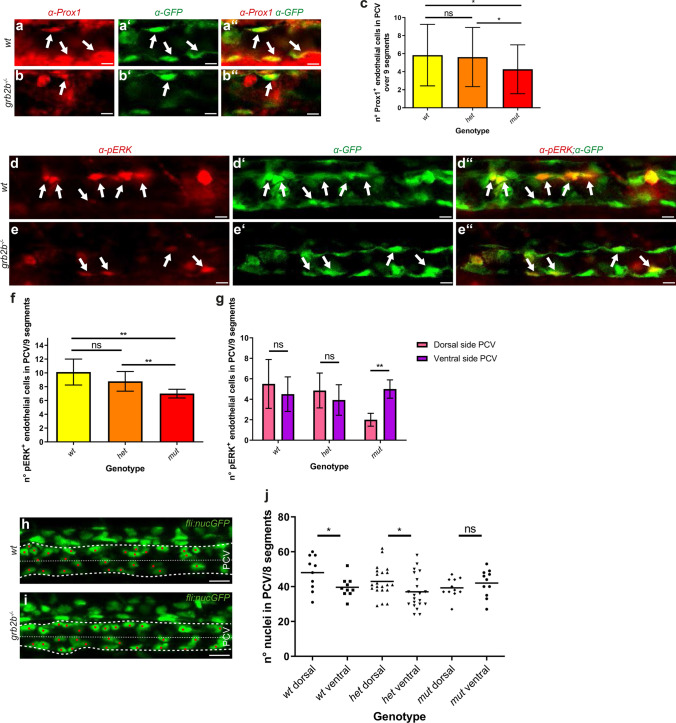
grb2b mutants show a defect in PCV polarization at 32hpf and exhibit decreased numbers of Prox1 and pERK positive endothelial cells. a–c The number of Prox1+ endothelial cells is significantly reduced in grb2bmu404 mutants at 32hpf. a–bʺ Confocal projections of the PCV at 32hpf in wild-type and mutant embryos. Prox1 antibody staining is shown in red and flt4:mCitrine in green, with arrows pointing at cells that co-express Prox1 and Flt4. c Quantification of Prox1+ endothelial cells in the PCV at 32hpf across 9 segments. wt: n = 38; het: n = 72; mut: n = 33. *Between wt and mut: P value = 0.0373 (t test, two-tailed); *Between het and mut: P value = 0.0412 (t test, two-tailed). d–f pERK+ endothelial cells are reduced in numbers in grb2bmu404 mutants at 32hpf. d–eʺ Confocal projections showing pERK antibody staining in red and endothelial cells in green (flt4:mCitrine) in the PCV at 32hpf. Arrows point at cells that express both pERK and Flt4. f Quantification of the number of pERK+ cells across 9 segments, demonstrating a decreased number in grb2b mutants. wt: n = 8; het: n = 14; mut: n = 6. **Between wt and mut: P value = 0.0013 (Mann–Whitney); ** between het and mut: P value 0.0036 (Mann–Whitney). g Moreover, pERK+ cells in wild type and heterozygotes are equally distributed between dorsal and ventral hemispheres of the PCV, whereas in mutants they are more concentrated in the ventral side. wt: n = 8; het: n = 14; mut: n = 6. **Between dorsal side and ventral side in mut: P value = 0.0022. h, i Confocal projections of fli:nEGFP wild-type and mutant embryos, highlighting the PCV between the more prominent dashed lines. The thinner dashed lines divide the PCV in two equal parts: the dorsal and the ventral side. j Quantifications of nuclei in the PCV at 32hpf showing that in grb2bmu404 mutants, endothelial cells are equally distributed between the ventral and the dorsal side. In siblings, endothelial cells are enriched in the dorsal part of the PCV. wt: n = 10; het: n = 21; mut: n = 11. *Between wt dorsal and wt ventral: P value 0.0327 (t test, two-tailed); *Between het dorsal and het ventral: P value = 0.044 (t test, two-tailed). ns not significant. Scale bars: a–bʺ: 10 µm; d–eʺ: 10 µm; h, i 20 µm. Data in c, f, g are mean ± s.d. Data in j represent the mean with individual data points. All quantifications have been done blindly

