Fig. 8.
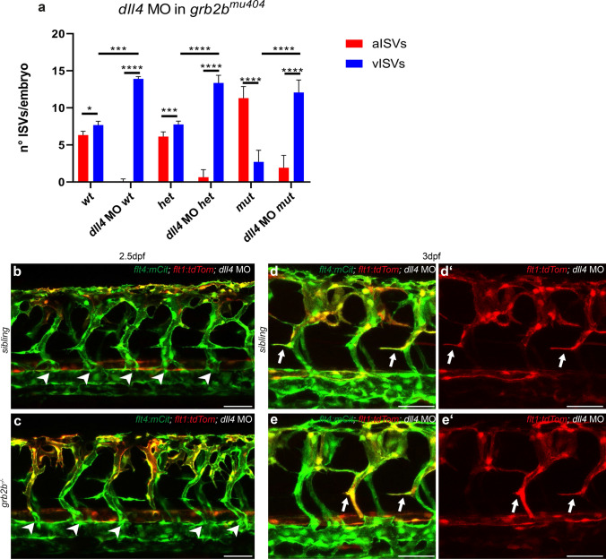
grb2b mutants show an increase in vISVs upon dll4 knockdown. a Quantifications of aISVs and vISVS at 2.5dpf in un-injected control and dll4 MO injected embryos from grb2bmu404 heterozygous parents. 14 ISVs per embryo were analyzed. *Between aISVs and vISV in wt: P value 0.013 (Mann–Whitney); ****Between aISVs and vISV in dll4 MO wt: P value < 0.0001 (Mann–Whitney); ***Between aISVs and vISV in het: P value 0.0009 (Mann–Whitney); ****Between aISVs and vISV in dll4 MO het: P value < 0.0001 (Mann–Whitney); ****Between aISVs and vISV in mut: P value < 0.0001 (Mann–Whitney); ****Between aISVs and vISV in dll4 MO mut: P value < 0.0001 (Mann–Whitney); ***Between vISVs wt and vISVs dll4 MO wt: P value = 0.0001 (Mann–Whitney); ****Between vISVs het and vISV in dll4 MO het: P value < 0.0001 (Mann–Whitney); ****Between vISVs mut and vISV in dll4 MO mut: P value < 0.0001 (Mann–Whitney). b–eʹ Confocal pictures of grb2b mutant or sibling embryos at 2.5dpf (b, c) and 3dpf (d–eʹ), injected with dll4 MO. Veins are shown in green (flt4:mCitrine) and arteries in red (flt1:tdTomato). Arrowheads highlight vISVs while arrows mark protrusions from ISVs extending towards the HM. aISV arterial intersegmental vessel, vISV venous intersegmental vessel, MO morpholino. Scale bars: 50 µm. Data in a represent mean ± sd

