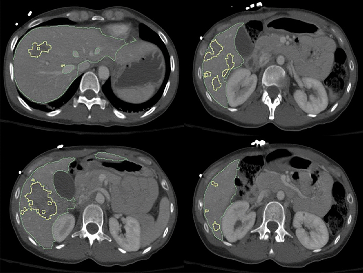
Figure 1.
Figure demonstrates high fidelity of deep learning-based segmentation of both the liver (green contour) and fine irregular margins of multifocal hepatic lacerations with variable shapes and sizes (yellow contour). The LPDI is automatically calculated as % liver disruption over total liver volume and was 13.1% in this example. (LPDI- liver parenchymal disruption index)

