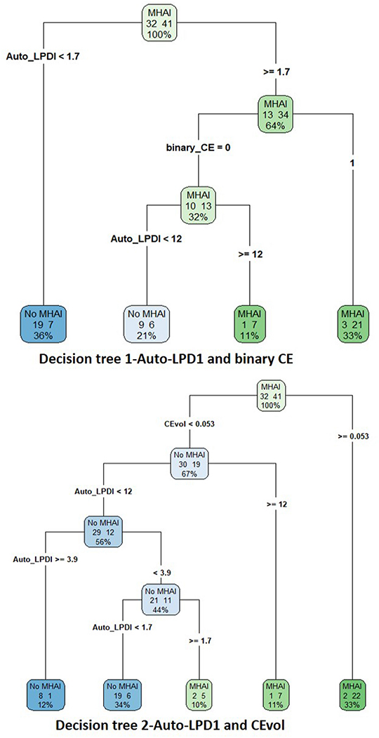
Figure 2.
Decision trees from CART analysis are shown. Decision tree 1 includes the combination of auto-LPDI and assessment of CE as a binary sign. Decision tree 2 includes auto-LPDI and CEvol. Contingency table results and AUCs for the two analyses is presented in Table 3. Decision tree analysis revealed an optimal cut off of ≥ 12% for ruling in MHAI when CE is either absent (decision tree 1), or diminutive at < 0.05 mL (decision tree 2). (CART-classification and regression trees, auto-LPDI- automated liver parenchymal disruption index, CE- contrast extravasation, CEvol- contrast extravasation volume)

