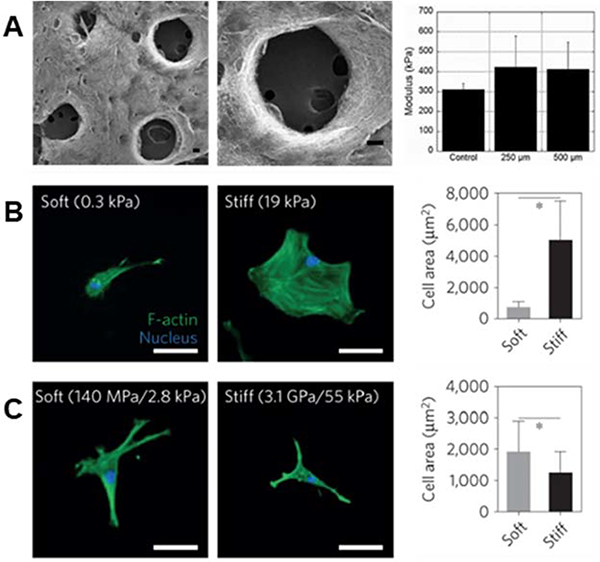
Figure 3. Importance of fiber physical properties for cell culture.
(A, left to right): SEM micrographs of MeHA fibers with user-specified photopatterned pores, zoomed in micrograph of a photopatterned pore, and a column chart displaying modulus of scaffolds – with no significant difference between scaffolds with pores and scaffolds without pores. (A) Reprinted and adapted with permission from Sundararaghavan et al., copyright 2010 John Wiley and Sons122; scalebars = 100 μm. (B, left to right): hMSCs show increased cell spreading on stiff hydrogels as opposed to soft hydrogels – quantified by the column chart illustrating cell area (*p < 0.05). (C, left to right): hMSCs demonstrate increased spreading on soft rather than stiff hydrogel fibers – quantified by the column chart showing cell area (*p < 0.05). These differing results emphasize the need for careful consideration when designing the biophysical properties of fibrous hydrogels for cell culture. (B) and (C) Reprinted and adapted with permission from Baker et al., copyright 2015 Springer Nature2; scalebars = 50 μm.

