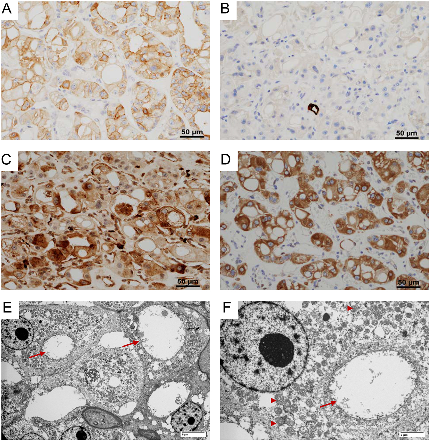
Figure 2. Immunohistochemical features of EVT.

EVT characteristically have (A) positive membranous CD117; (B) negative cytokeratin 7; (C) focal positive cathepsin-K; and (D) diffuse phospho-S6 expression. (E-F) Ultramicrographs showing numerous mitochondria (arrow head) and dilated cisterns of rough endoplasmic reticulum (arrow).
