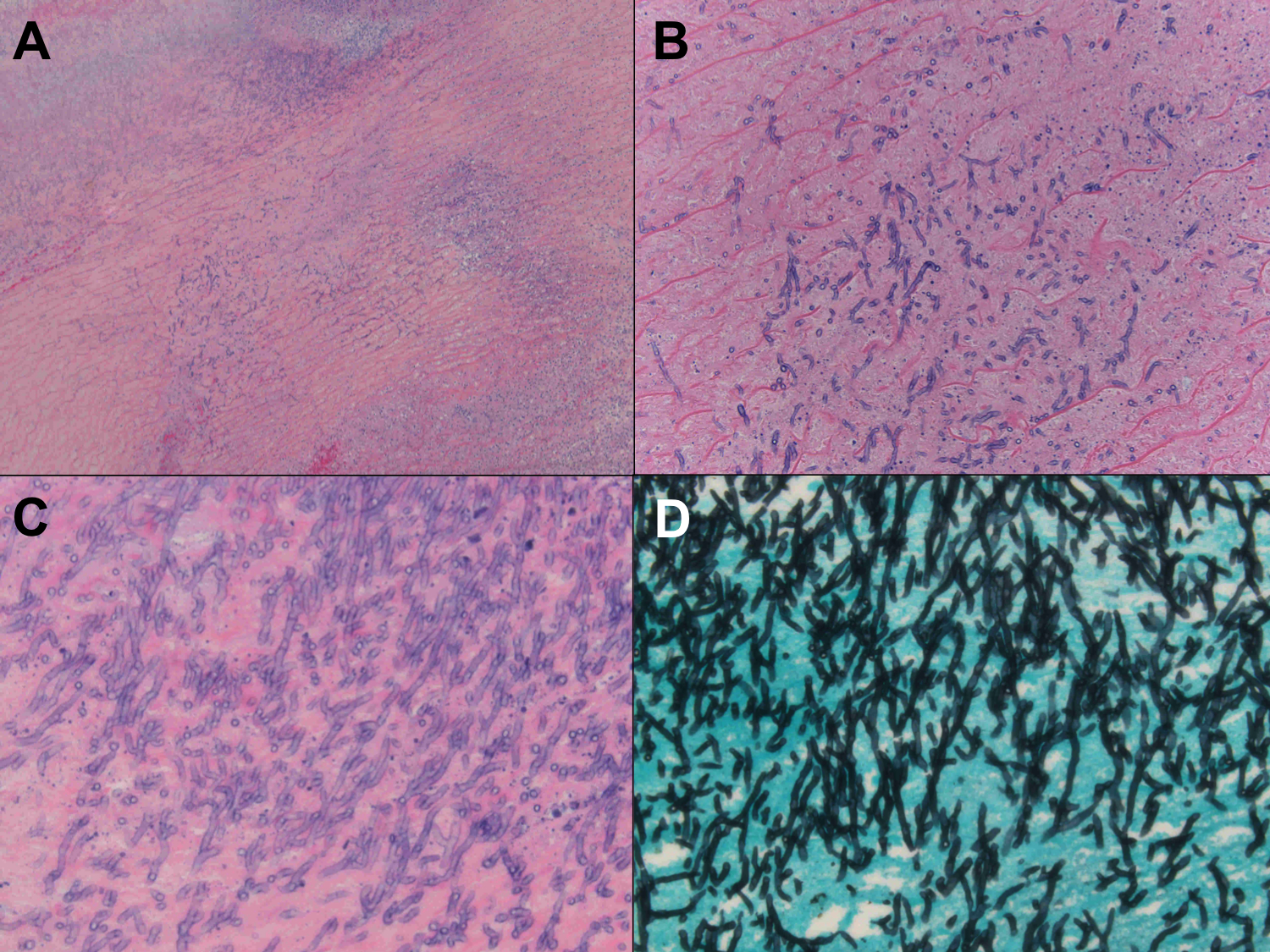
FIGURE 2. Pathology from aortic tissue excision demonstrating Aspergillus fumigatus.

(A) Hematoxylin and eosin (H&E) image (5x) demonstrates mostly necrotic vessel wall. (B) H&E image (20x) reveals nonviable vessel wall with fungal hyphae. (C) Fungal hyphae with acute angle branching shown on H&E (40x) image. (D) Grocott special stain (40x) highlights numerous fungal hyphae with acute angle branching.
