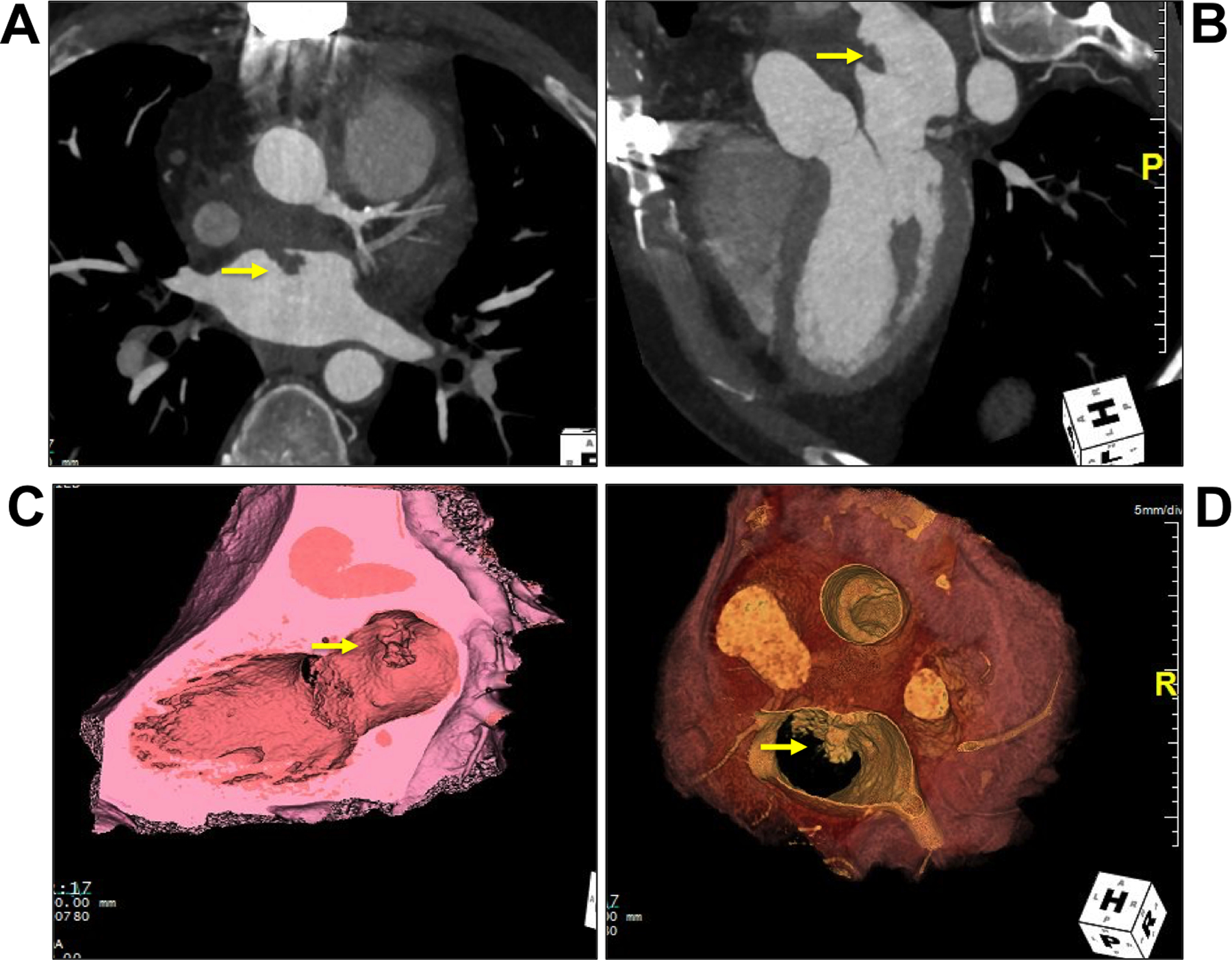
FIGURE 3. Selected images from follow-up contrast enhanced chest computed tomography.

Axial image (A), coronal oblique image (B), and long axis (C) and short axis (D) 3D endoluminal reconstruction demonstrates a large mass posterior to the ascending aorta graft that protrudes into the left atrium (arrow).
