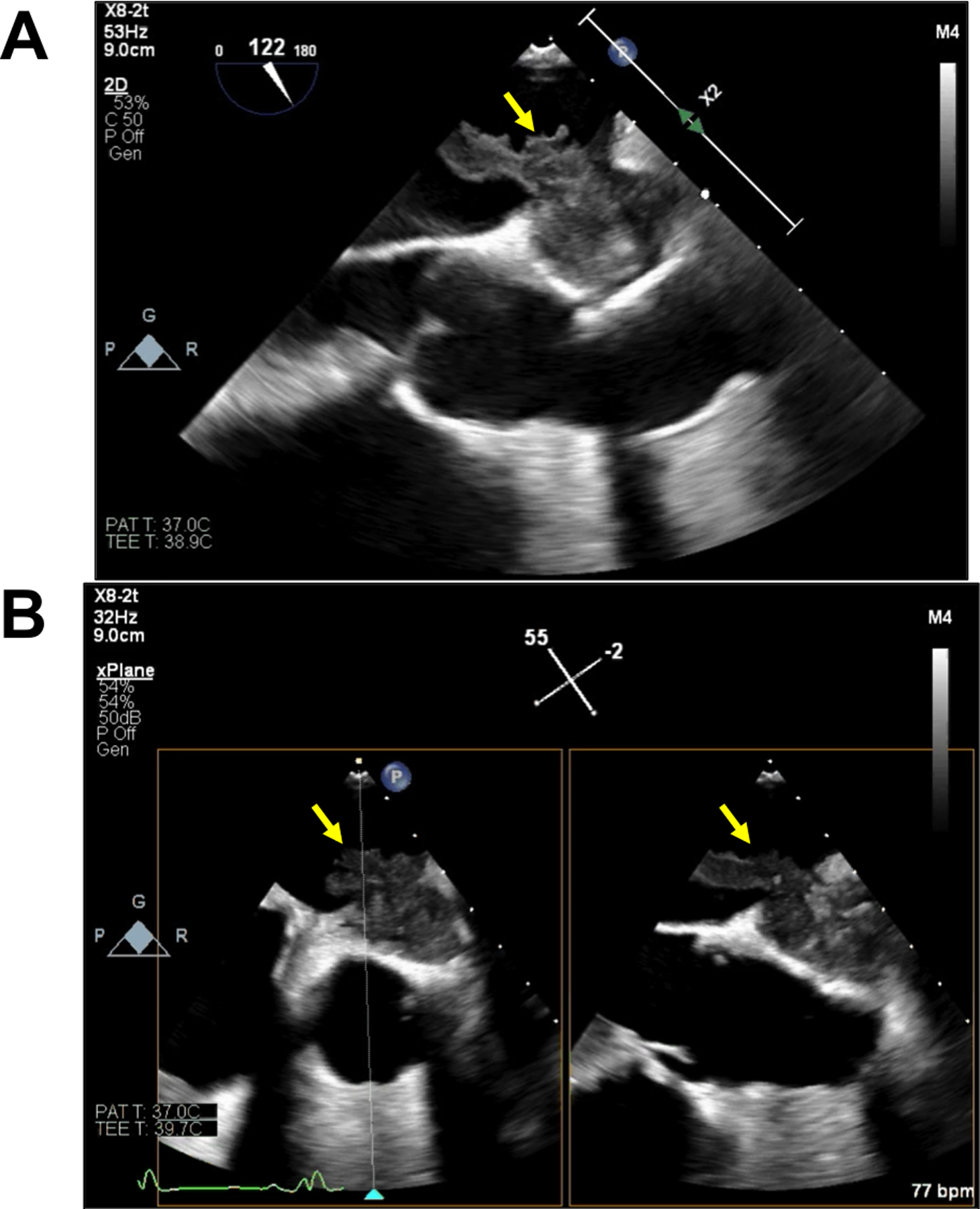
FIGURE 4. Selected images from transesophageal echocardiography.

Mid-esophageal ascending aorta long axis view (A) and x-plane view (B) reveal a large echogenic mass posterior to the ascending aorta graft invading the anterior left atrial wall with a large, irregular mobile component (2.6 cm × 1.0 cm) extending into the left atrial cavity.
