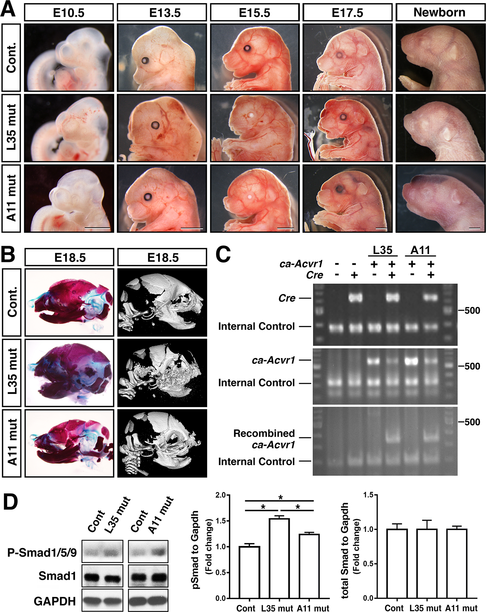
FIGURE 2.

Neural-crest specific augmentation of BMP signaling via ACVR1-Q207D develops distinctive phenotypes in two transgenic lines. A: Lateral views of the head region of control and two transgenic lines during mid to late gestation, and newborn stages. Bar =1 mm. B: Assessment of skeletal abnormalities at E18.5 through Alcian blue/alizarin red staining (left) and microCT (right). C: Genomic PCR to confirm genotypes of each embryos. Primers used; Cre F and Cre R (top), TF41 and TF61 (middle), and TF41 and CJ-Green (bottom) to detect presence of Cre, ca-Acvr1, recombined ca-Acvr1, respectively. D: Protein lysates were prepared from E10.5 embryos and BMP-Smad levels were measured by western blots using antibodies against total Smad1 and pSmad1/5/9. Levels of signals were normalized by GAPDH. *, p<0.05. L35 mut, ca-Avcr1 line 35 embryos (Fukuda, et al., 2006) also carrying P0-Cre transgene. A11 mut, ca-Avcr1 line A11 (this study) embryos also carrying P0-Cre transgene.
