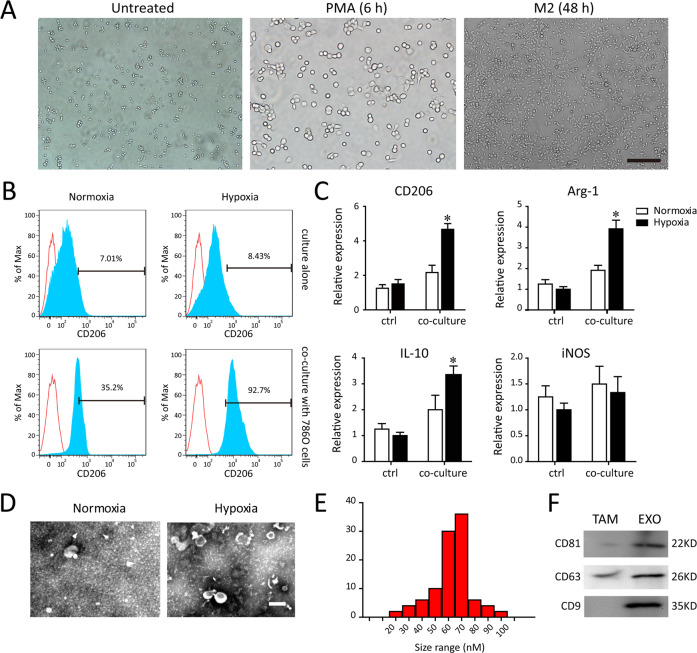
Fig. 1. Characterization of TAMs-derived exosomes.
A After PMA stimulated-differentiation, round and floating THP-1 cells became adherent flattened cells. B Flow cytometry analysis for the percentage of M2 polarized macrophages (CD206) after exposing to normoxia or hypoxia and co-culturing with or without 786-0 cells. C The RT–PCR detection of typical M2 markers (CD206, Arg1, IL-10, iNOS) in macrophages after exposing to normoxia or hypoxia and co-culturing with or without 786-0 cells. D Representative TEM images of TAM-derived exosomes (TAM-exo). Scale bar = 100 nm. E Size distributions of exosome fractions isolated from TAM conditioned medium by nanoparticle tracking analysis. F Western blot analysis of the exosomal markers (CD81, CD9, and CD63). *P < 0.01 vs. normoxia group.

