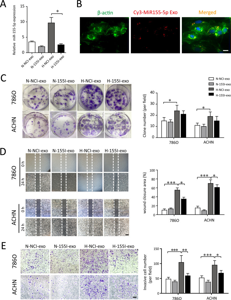
Fig. 4. Exosomal miR-155-5p facilitates the proliferation, wound healing and migration of RCC cells in vitro.
A The normoxic or hypoxic TAMs were transfected with antagomir control and antagomiR-155-5p to produce NCI-exos and 155I-exos. The corresponding exsomal miR-155-5p levels (N-exo+agomir-NC, N-exo+agomiR-155-5p, H-exo+antagomir-NC, H-exo+antagomiR-155-5p) were validated by RT–PCR. B The uptake of the Cy3-labeled M2-Exos was observed in RCC cells. C 786-0 and ACHN cells were incubated with exosomes derived from the normoxic or hypoxic TAMs, which were transfected with control or agomiR-155-5p, respectively. Clone formation of each group (N-NCI-exo, N-155I-exos, H-NCI-exo, H-155I-exo) were observed and the colony numbers were quantified. D The RCC cells were scratched, photographed at time 0, and incubated in the presence of N-NCI-exo, N-155I-exos, H-NCI-exo, H-155I-exo (50 μg/ml). Photographs were taken again after 24 h (Scale bar = 200 μm). Quantification of the closure area was presented as the ratio of closure area to the initial wound area. E Representative image of invasion assay. Diagram of invasive cells in response to various exosomes from six random high-power fields (Scale bar = 100 μm) counted in three independent experiments. *P < 0.05, **P < 0.01, ***P < 0.001.

