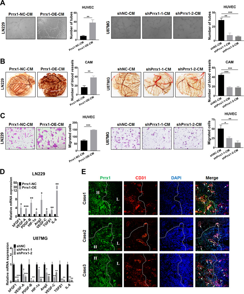
Fig. 3. Prrx1 promotes glioma angiogenesis through upregulating proangiogenic factors in NSTCs.
A Representative capillary tubule structures were shown for HUVECs treated with culture medium collected from the indicated LN229 and U87MG cells. Scale bar represents 50 μm. B Blood vessels formed in representative images of the CAM assay after CM treatment. C Transwell assay was performed in HUVECs to detect the effect of CM treatment on cell migration. Scale bar represents 50 μm. D RT-qPCR detects the effects of Prrx1 on the expression of classical proangiogenic factors in U87MG and LN229 cells (mean ± SD, n = 3). E IF assay analysis of glioma specimens showed the vessel density in regions with different Prrx1 expression levels. The H indicated regions with high expression level of Prrx1 and the L indicated regions with low Prrx1 expression. Representative images of three cases were shown. Scale bar represents 10 μm. *P < 0.05, **P < 0.01, ***P < 0.001.

