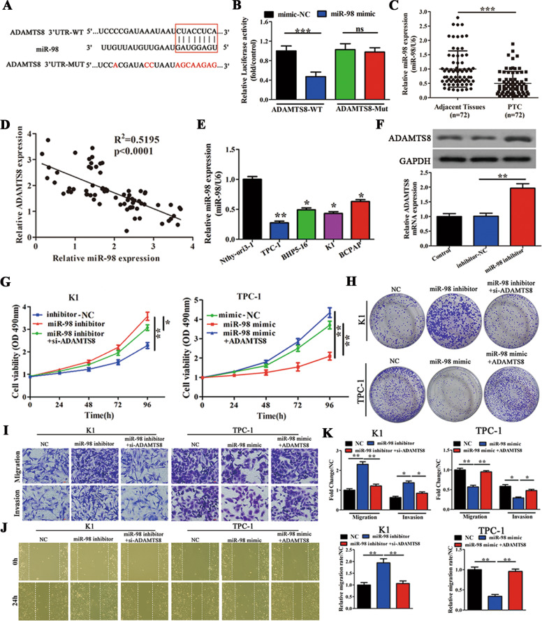
Fig. 5. ADAMTS8 is a direct target of miR-98 and is involved in miR-98-mediated cell proliferation and migration/invasion.
A WT 3′-UTR of ADAMTS8 (ADAMTS8 3′-UTR-WT) and a mutant 3′-UTR of ADAMTS8 with mutations at the predicted miR-98 binding site (ADAMTS8 3′-UTR-MUT) (n = 3). B A luciferase reporter vector carrying ADAMTS8 3′-UTR-WT or ADAMTS8 3′-UTR-MUT (or the empty vector) was co-transfected with miR-98 mimic or mimic-NC, and relative luciferase activity was measured 48 h post transfection (n = 3). C qRT-PCR analysis of miR-98 expression in 72 paired PTC tissues and corresponding adjacent tissues. D Pearson correction of miR-98 and ADAMTS8 were analysed (n = 72). E qRT-PCR analysis of miR-98 expression in four PTC cell lines (TPC-1, BHP5-16, K1, and BCPAP), and in Nthy-ori3-1 cells (n = 3). F Western blot and qRT-PCR analyses of ADAMTS8 expression levels in K1 cells transfected with inhibitor-NC or miR-98 inhibitor (n = 3). G, H MTT and colony-forming growth assays were performed to determine the proliferation of K1 and TPC-1 cells (n = 3). I Transwell assays were performed to determine the migration and invasion capacity of K1 and TPC-1 cells (n = 3). Scale bars = 50 μm. J Wound healing assays were performed to assess the migratory capacity of K1 and TPC-1 cells (n = 3). K The migration and invasion abilities and the migratory activity (wound healing) were calculated and compared to the different vectors (n = 3). *p < 0.05, **p < 0.01, and ***p < 0.001.

