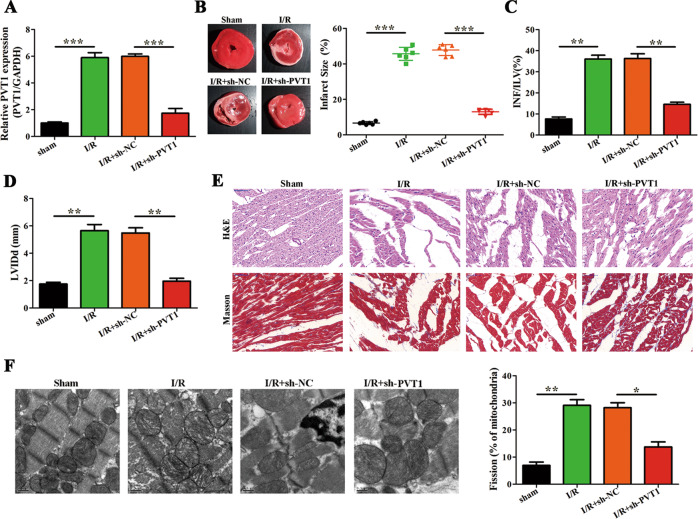
Fig. 4. LncRNA PVT1 promotes myocardial I/R injury and mitochondrial fusion and fission in vivo.
A sh-PVT1 or sh-NC was intraperitoneally administered 24 h before myocardial I/R injury (n = 6 per group). Post I/R injury, RNA was isolated from heart tissues and expression of PVT1 was determined by qRT-PCR. Relative fold change of PVT1 expression with respect to GAPDH is shown (n = 6). B–D Quantification of I/R injury. Representative images of heart tissues in the left panel for different groups are shown. Other panels indicate infarct size (%), INF/LV (%), and LVIDd (mm) (n = 6). E Representative images of H&E and Masson’s trichrome staining of left ventricle [43] sections (Scale bar = 50 μm) (n = 6). F Representative TEM images of heart tissues are shown in the left panel. Quantification of fragmented mitochondria is shown in the right panel (Scale bar = 0.5 μm) (n = 6). *P < 0.05; **P < 0.01; ***P < 0.001.

