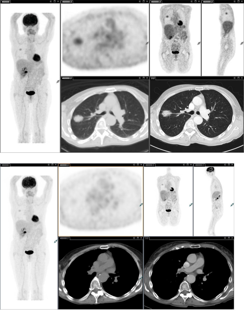
Fig. (2).
Moderately differentiated adenocarcinoma G2 (70% Acinar, 20% Lepidic, 10% Papillary) of the upper lobe of the right lung (SUVmax 3.5). Surgery identified hilar node metastasis (SUVmax 2.0; SUVmax liver 2.5) not identified by FDG PET/CT Imaging. It should be kept in mind that in patients with less avid primary tumors, node metastases may be missed by FDG PET/CT imaging. (A higher resolution / colour version of this figure is available in the electronic copy of the article).

