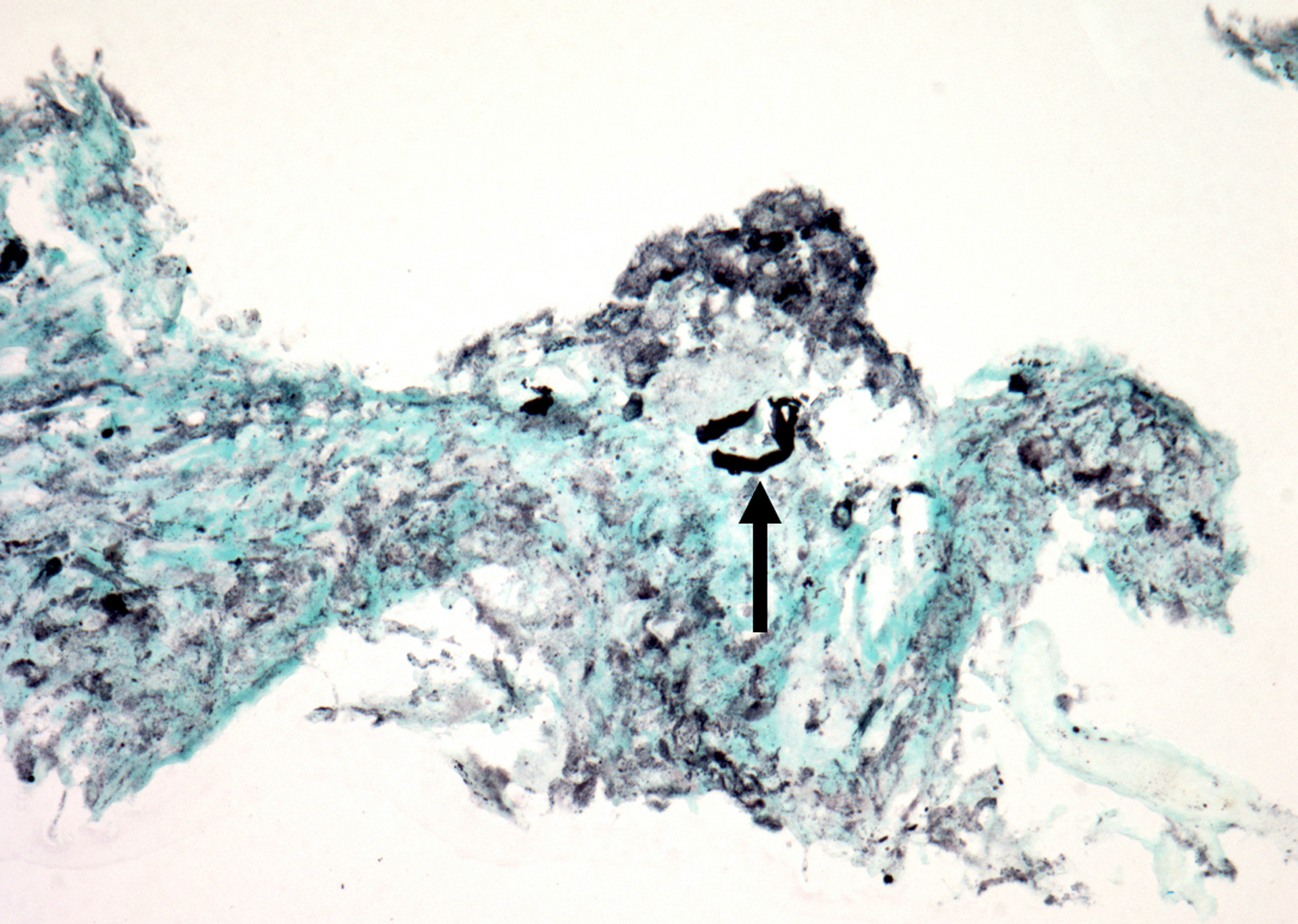
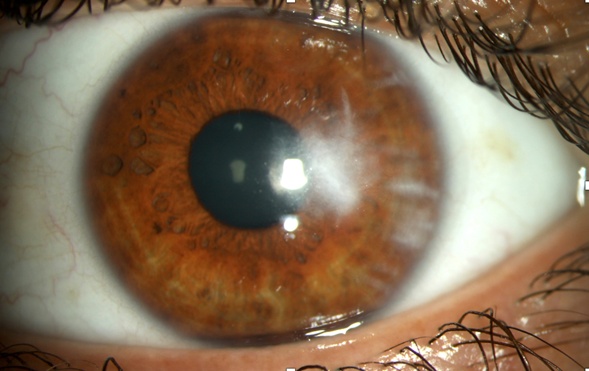
Figure 3.


Histopathology photographs of the surgically removed endothelial plaque and slit lamp photograph of the patient’s left eye 4 weeks following the procedure. (A) Stains and cultures of the removed specimen were negative, but histopathology revealed fungal elements (arrow) within ulcerated corneal stroma (Gomori-methenamine silver; original magnification × 400). (B) The patient experienced transient focal edema at the site of the biopsy, which resolved entirely by the fourth postoperative week. A deep stromal scar remained in the area and the patient’s vision was restored to 20/20.
