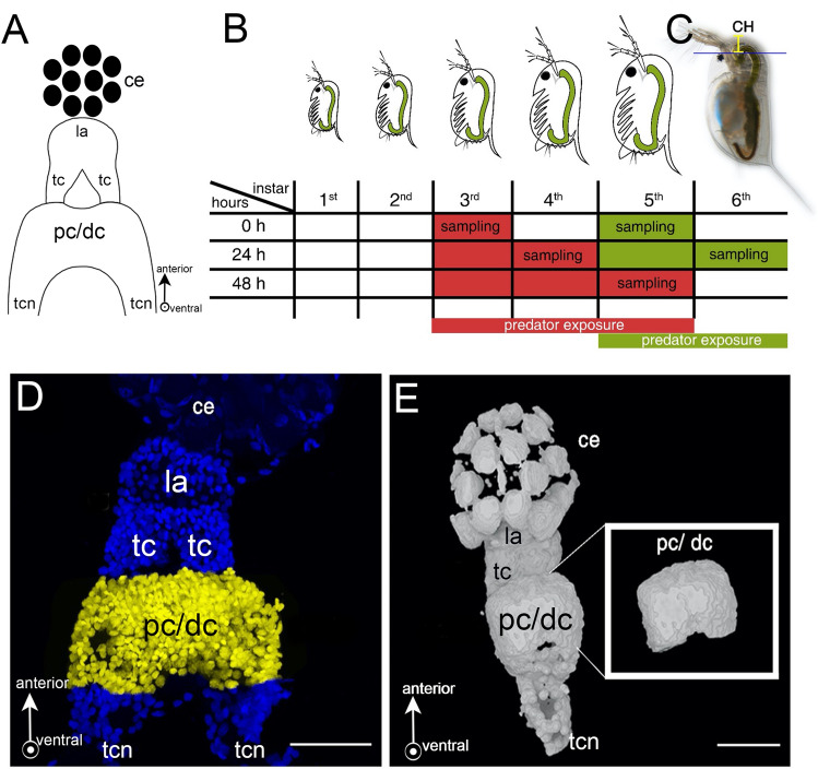
Figure 1.
Schematic overview of experimental procedures. (A) Schematic frontal view of a Daphnia brain displaying the optic ganglia: composed of lamina and tecta, and pc/dc-complex. (B) Points in time of D. longicephala induction. Predator exposure started in the 3rd or 5th instar, respectively. When exposed in the 3rd instar, individuals were sampled at 0 h, 24 h and 48 h. To validate our findings in a different instar, animals were also exposed in the 5th instar; here we focussed on the target stages i.e. 0 h and 24 h. Controls were performed alongside the respective treatment but without predators. (C) Measurement of morphological defence parameters. We measured the crest height as the distance between the upper eye margin and the rostral extension of the head (yellow line). (D) Dissected brain mount stained with DAPI. We counted the cells of the pc/dc-complex (yellow). (E) 3D projection of a brain mount from confocal stacks for volumetrics created with MorphoGraphX Version 1.1.1280-CellAtlas (available from https://www.mpipz.mpg.de/MorphoGraphX/software). Only the pc/dc-complex (insert) was used to determine the brain volumes. (A, D, E) Arrows depict the anterior and ventral (arrow points out of the drawing pane) orientation. Abbreviations: CH, crest height; ce, compound eye; la, lamina; tc: tecta, pc/dc, pc/dc-complex; tcn, tritocerbral neuropil.

