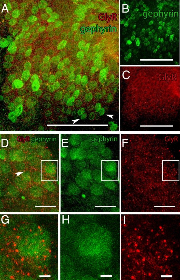
Figure 4.
Co-localization of gephyrin and glycine receptor (GlyR). (A) Merged display of anti-gephyrin and anti-GlyR staining pattern. Both antibodies are identified within the pc/dc-complex. Anti-GlyR staining colocalized with the anti-gephyrin antibody staining. Not all gephyrin-labelled cells show markings of anti-GlyR (white arrow heads). (B) Gephyrin is found in the cell soma and on the cell surface evenly distributed within the pc/dc-complex. (C) GlyRs are evenly distributed in the pc/dc-complex and the staining pattern shows the typical receptor spots. A–C: Scale bar 50 µm. (D) Merged display of anti-gephyrin and anti-GlyR staining on the cellular level. Anti-gephyrin staining is found throughout the whole cytoplasm, while anti-GlyR staining is limited to concentrated spots on the cell surface (double arrow heads). Inset is magnified in G. (E) Staining pattern of gephyrin on the cellular level displayed in green. Inset is magnified in H. (F) Staining pattern of anti-GlyR on the cellular level displayed in red. Inset is magnified in I. D–F: Scale bar 10 µm. (G) Magnifying inset (displayed in D) of the concentrated Glycin receptors (red) in colocalization with gephyrin labelled cells (green). (H) Magnifying inset displayed in (E) of gephyrin labelled cells (green). (I) Magnifying inset (displayed in F) of concentrated Glycin receptors (red). G–H: Scale bar 5 µm. Images were created with ImageJ 1.52n (Fiji, available from https://fiji.sc/)38.

