Figure 1.
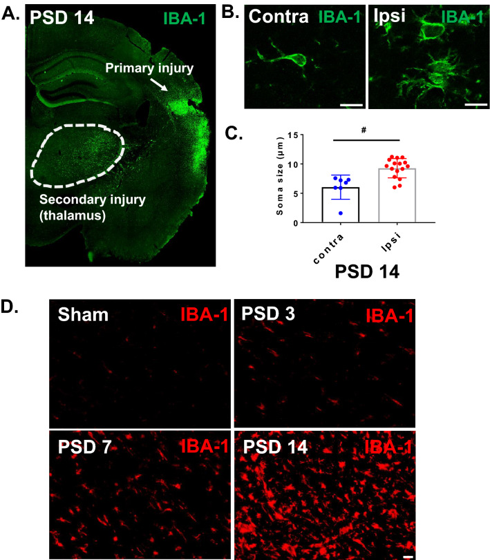
Cortical stroke induces delayed microglial activation in the thalamus. Cortical infarction was induced in young C57BL/6 J mice. At 3 days, 1 week and 2 weeks after stroke, brains were isolated and sectioned. Immunostaining with anti-IBA-1 antibody was performed. Brain sections at − 2 mm from bregma were used for the visualization of thalamic microglial activation. (A) Coronal brain section showing activated MG/macrophages (IBA-1) in the posterior region of the primary injury and in the ipsilateral thalamus at PSD 14 (10X obj, stitched image). (B) Enlarged images showing morphology of MG/macrophages in the ipsilateral and contralateral thalamus at PSD 14 (scale bar = 10 µm, 63X obj). (C) The microglial soma size, located in the ipsilateral thalamus, is much bigger than microglia in the contralateral thalamus (# p < 0.05, unpaired t test) (D) Time course of IBA-1 staining showing delayed MG/macrophage activation in the ipsilateral thalamus (scale bar = 20 µm).

