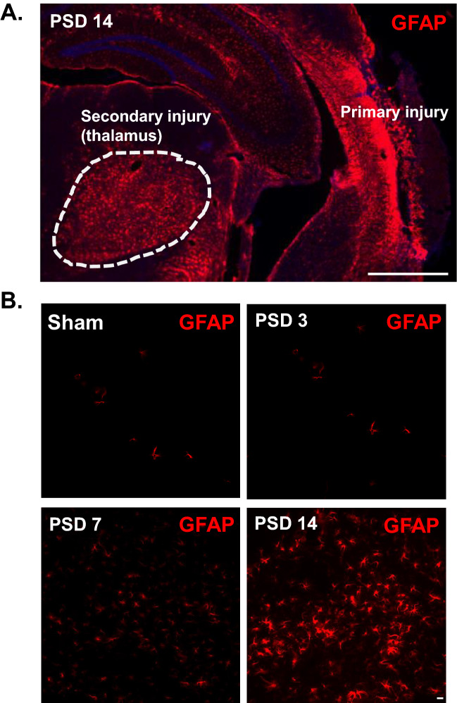
Figure 4.
Cortical stroke induces delayed astrogliosis in the ipsilateral thalamus. Cortical infarction was induced in young C57BL/6 J mice. At 3 days, 1 week and 2 weeks after stroke, brains were isolated and sectioned. Immunostaining with anti-GFAP antibody was performed. Brain sections at − 2 mm from bregma were used for the visualization of thalamic astrocyte activation. (A) Coronal brain section showing activated astrocytes (GFAP) in the posterior region of the primary injury and in the ipsilateral thalamus at PSD 14. A glial scar is evident in the cortical injury, but not within the thalamus (10X obj, stitched image; scale bar = 1 mm). (B) Time course of GFAP staining showing delayed astrocyte activation in the ipsilateral thalamus (20X obj; scale bar = 20 µm).

