Figure 5.
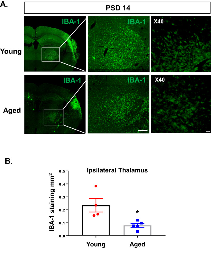
Aged mice demonstrate reduced IBA-1 expression in ipsilateral thalamus at PSD 14 compared with young mice. (A) Representative images comparing IBA-1expression in brain of young (11–13 weeks) and aged (19–21 months) mice at PSD 14. Imaged with 10X obj, stitched image; scale bar = 1 mm (left panel) or 250 µm (middle panel). Blow up imaged with 40X obj; scale bar = 20 µm (right panel). (B) Quantification of total IBA-1 stained area in ipsilateral thalamus of young and aged mice. * P < 0.05, unpaired t test.

