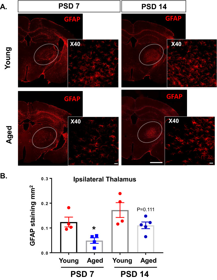
Figure 6.
Aged mice demonstrate reduced astrogliosis in ipsilateral thalamus at PSD 7 compared with young mice. GFAP expression was evaluated at PSD 7 and PSD 14 in ipsilateral thalamus from young and aged mice. (A) Representative images showing GFAP expression in coronal sections at -2 mm from bregma (10X obj, stitched image; scale bar = 1 mm). Insert images was taken at 40X, scale bar = 20 µm. (B) Quantification of total GFAP stained area in ipsilateral thalamus of young and aged mice. * P < 0.05, unpaired t test. (P = 0.111 for young versus aged at PSD 14).

