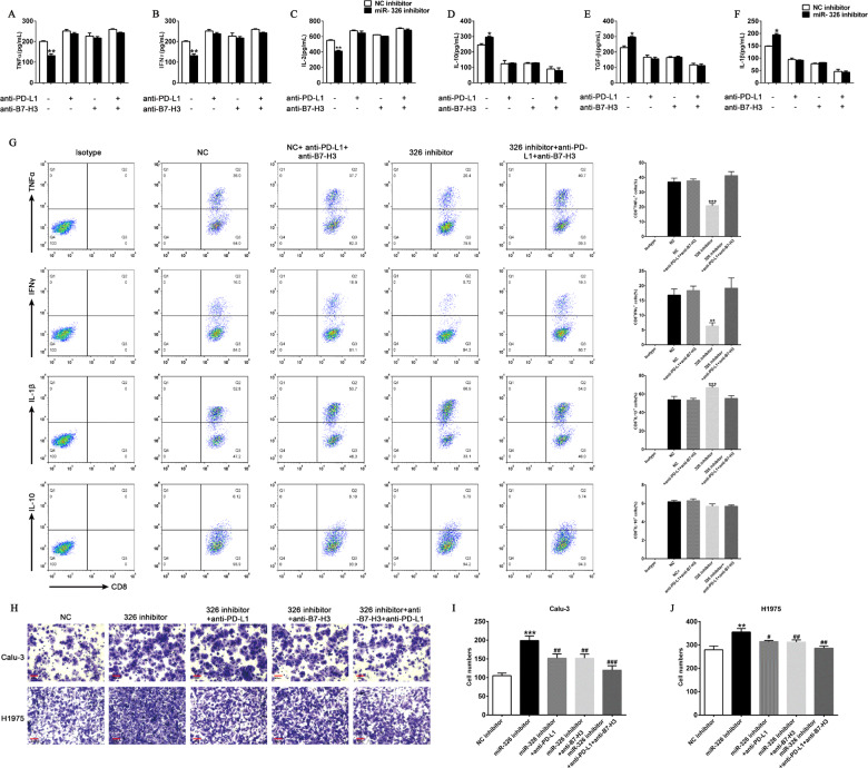
Fig. 5. Blockade of PD-L1 and B7-H3 markedly abrogates the aggressive tumor behavior caused by miR-326 downregulation.
A–F T cells were co-cultured with miR-NC- or miR-326-overexpressed H1975 cells, and PD-L1/B7-H3 blocking antibody was added. Cytokines in the supernatant were measured by ELISA (n = 3). G TNF-α, IFN-γ, IL-10, and IL-1β expression of CD8+ T cells after miR-326 overexpression and/or PD-L1/B7-H3 antibody incubation by flow cytometry (n = 3). H Transwell assay of control and miR-326-overexpressed cells, and PD-L1/B7-H3 blocking antibody was added. I, J Histogram of the number of migrated cells (n = 3). All data were presented as mean ± SEM. Comparisons between groups for statistical significance were performed with one-way (I, J) or two-way (A–G) ANOVA with Tukey’s post hoc test. *P < 0.05, **P < 0.01, ***P < 0.001 versus NC inhibitor. ##P < 0.01, ###P < 0.001 versus miR-326 inhibitor. Scale bar, 200 µM.

