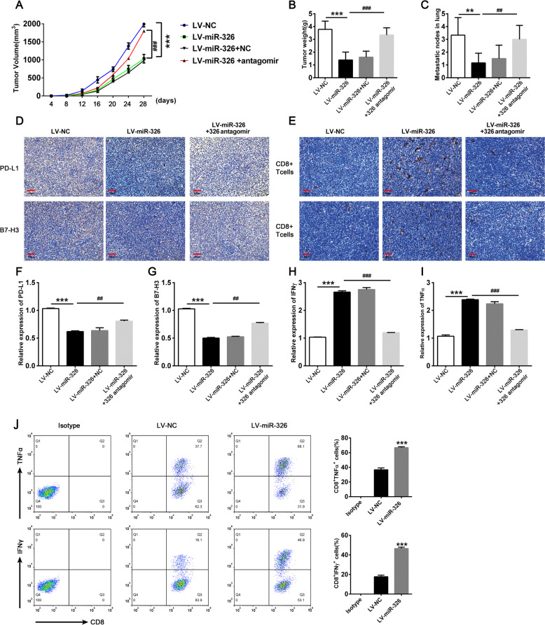
Fig. 6. Overexpression of miR-326 enhances TNF-α and IFN-γ expression of infiltrated CD8+ T cells and prevents metastasis by targeting PD-L1 and B7-H3 on cancer cells.
5 × 10⁵ miR-326 overexpressed LLC cells or empty vector control LLC cells were subcutaneously injected into C57BL/6 mice. MiR-326 antagomir or NC antagomir were intratumor injected when the tumor diameter reached 0.5 cm. The mice were sacrificed 28 days after inoculation. A Tumor volume (n = 7). B Tumor weight (n = 6). C Pulmonary metastasis nodule calculated by hematoxylin–eosin staining. (n = 6) D, E PD-L1, B7-H3 expression, and CD8+ T cells infiltration were determined by IHC staining in tumor tissues. F–I PD-L1, B7-H3, TNF-α, and IFN-γ were detected by qRT-PCR in tumor sites (n = 3). J TNF-α and IFN-γ expression of infiltrated CD8+ T cells by flow cytometry (n = 3). All data were presented as mean ± SEM. Comparisons between groups for statistical significance were performed with one-way (B, C and F–I) or two-way (A) ANOVA with Tukey’s post hoc test or Student’s t test (J). *P < 0.05, ***P < 0.001 versus LV-NC. #P < 0.05, ##P < 0.01, ###P < 0.001 versus LV-miR-326. Scale bar, 200 µM.

