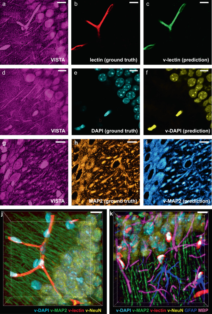
Fig. 4. Label-free VISTA prediction for specific and multi-component imaging of brain hippocampal tissues.
a–c The input VISTA image (a), the ground truth fluorescence image of lectin-DyLight594 stained blood vessels (b), and the predicted VISTA-lectin (v-lectin) image of blood vessels (c). d–f The input VISTA image (d), the ground truth fluorescence image of DAPI stained nuclei (e), and the predicted VISTA-DAPI (v-DAPI) image of nuclei (f). g–i The input VISTA image (g), the ground truth immunofluorescence image of MAP2 stained neuronal cell bodies and dendrites (h), and the predicted VISTA-MAP2 (v-MAP2) image of neuronal cells and dendrites (i). j Four-color volume imaging from label-free VISTA prediction for vessels (v-lectin, red), nuclei (v-DAPI, cyan), neuronal cell bodies, and dendrites (v-MAP2, green), and matured neuron cell bodies (v-NeuN, yellow). k Tandem 6-color volume imaging from label-free 4-color VISTA prediction and parallel two-color immuno-fluorescence images of GFAP (blue) and MBP (magenta). Scale bars: 40 μm. The length scale is in terms of distance after sample expansion.

