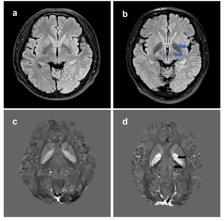
Fig. (2).
Brain MRI changes in WD. T2-FLAIR weighted images and QSM are used to show brain MRI changes in a 27-year-old WD patient. (a) Normal T2-FLAIR weighted images. (b) Hyperintensities in the bilateral globus pallidus and thalamus (blue arrows) can be observed in T2-FLAIR weighted images in the WD patient. (c) Normal QSM images. (d) QSM images show increased susceptibility (hyperintensities, black arrows) of bilateral globus pallidus and thalamus in the same patient. QSM, quantatitive susceptibility mapping; T2-FLAIR, T2-fluid attenuated inversion recovery; WD, Wilson’s disease. (A higher resolution / colour version of this figure is available in the electronic copy of the article).

