
Figure 1.
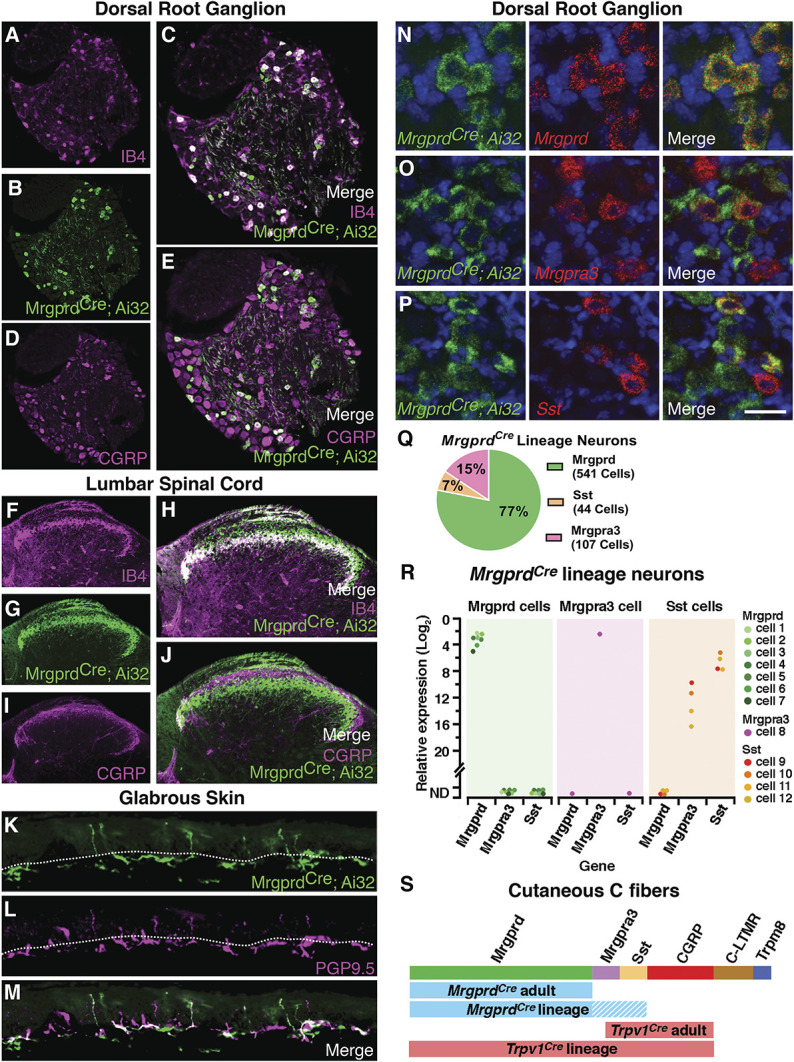
MrgprdCre lineage afferents, which comprise Mrgprd, Mrgpra3, and Sst populations, target lamina IIi in the dorsal horn and the superficial aspect of the skin. (A–E) Representative images of a lumbar dorsal root ganglion from a MrgprdCre; Ai32 mouse that is costained for eYFP and IB4 or CGRP. (F–J) Representative images of the superficial dorsal horn, lumbar level from a MrgprdCre; Ai32 mouse that is costained for eYFP and IB4 or CGRP. (K–M) Representative images of the glabrous skin from a MrgprdCre; Ai32 mouse that is costained for eYFP and PGP9.5. Dotted line demarcates the epidermal–dermal junction. (N–P) Representative RNA FISH images of lumbar dorsal root ganglion from a MrgprdCre; Ai32 mouse that is costained with probes against Eyfp, Mrgprd, Mrgpra3, and Sst. (Q) The relative proportion of primary afferent populations captured by the MrgprdCre lineage as determined by FISH. Data are from 3 mice, with 4 to 5 slices imaged per mouse for a total of 692 MrgprdCre lineage neurons counted per probe combination. (R) Expression of Mrgprd, Mrgpra3, and Sst mRNA relative to Gapdh in individual eYFP-marked MrgprdCre lineage neurons. Data are presented as the log2 ΔCT expression relative to Gapdh expression within the same cell such that smaller numbers represent higher mRNA expression. Colored dots represent data points from individual cells from a single, representative mouse. (S) Schematic to illustrate the different subtypes of cutaneous C fibers that are captured by the MrgprdCre and Trpv1Cre alleles with developmental (lineage) or adult recombination. FISH, fluorescence in situ hybridization.