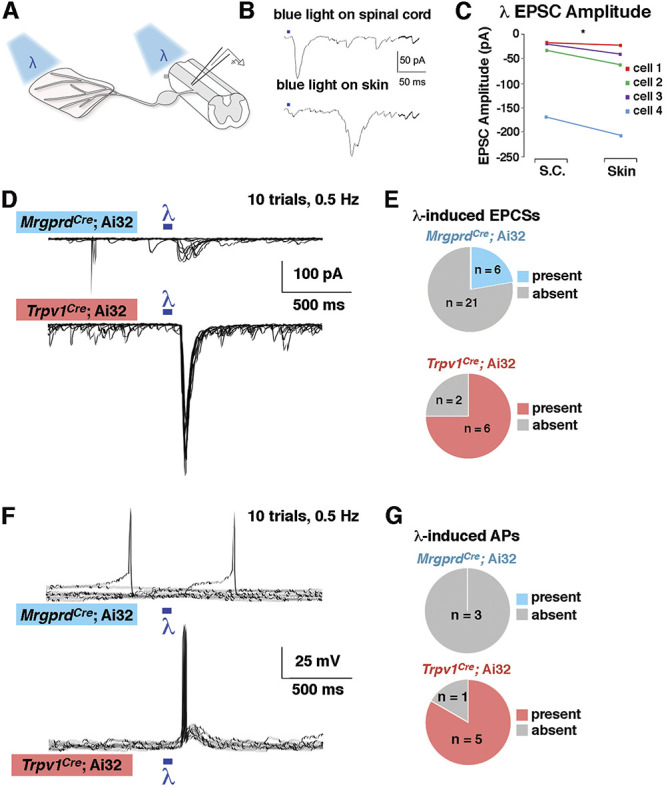
Figure 5.

Lamina I neurons receive strong input from Trpv1Cre lineage afferents but weak input from MrgprdCre lineage afferents (A) Schematic of recordings with optogenetic stimulation at either the peripheral or central terminal. (B–C) Representative traces and quantification of EPSC amplitude from lamina I neurons after optogenetic stimulation at either the skin or the spinal cord. *P < 0.05 (paired Student t test). (D and E) Representative voltage clamp recordings (VH = −70) and quantification of the proportion of lamina I neurons that show EPSCs on optogenetic stimulation at the spinal cord in MrgprdCre; Ai32 and Trpv1Cre; Ai32 mice. (F–G) Representative current clamp recordings and quantification of the proportion of lamina I neurons that show action potentials on optogenetic stimulation at the spinal cord in MrgprdCre; Ai32 and Trpv1Cre; Ai32 mice. For D and F, the responses to 10 stimulus presentations are superimposed. EPSC, excitatory postsynaptic currents.
