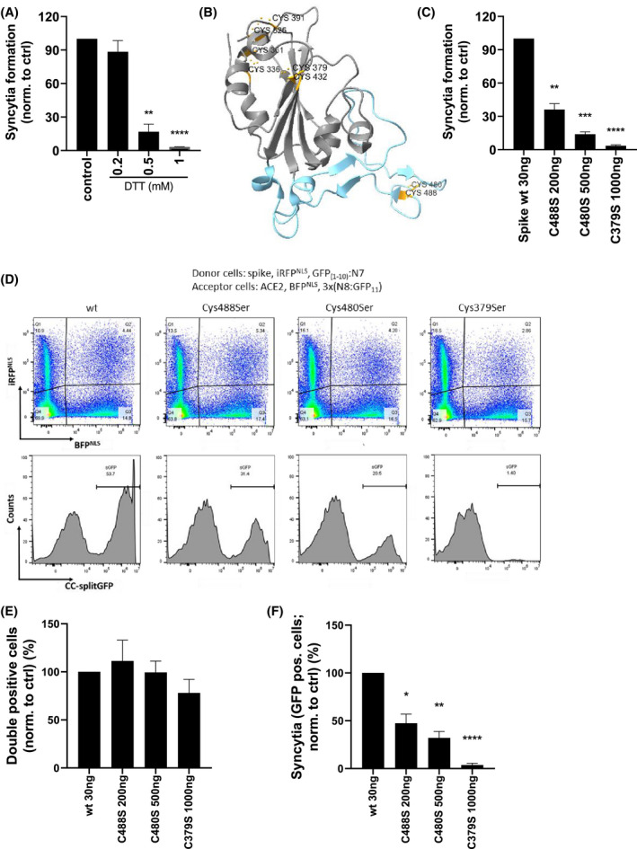
FIGURE 4.

Mutation of cysteine residues in RBM decreases the fusogenic ability of spike protein. A, HEK293T spike‐expressing cells were preincubated for 30 minutes with the indicated concentrations of DTT. ACE2‐expressing cells were added, mixed, and seeded. After 3 hours, cell fusion was detected by measuring luciferase activity. B, Structure of SARS‐CoV‐2 spike RBD with indicated cysteine pairs (orange) and RBM (blue). C, HEK239T were transfected with the indicated amounts of spike wt or cysteine mutants and cLuc:N7 or ACE2 and nLuc:N8 plasmids. After 24 hours, the cells were mixed and seeded. After 3 hours, cell fusion was detected by measuring luciferase activity. D‐F, HEK239T were transfected with the indicated amounts of spike wt or cysteine mutants, iRFPNLS and split GFP(1‐10):N7 or ACE2, BFPNLS, and split 3x(N8:GFP11) plasmids. The fusogenic ability of the spike was determined by syncytia formation after mixing with ACE2‐expressing cells and measuring on a flow cytometer after 3 hours. The percentage of double‐positive cells (E) and syncytia (F) normalized to spike wt‐expressing cells is shown. Combined means from three (A, C) or pooled data from four (E, F) independent experiments are shown as mean ± SEM P values of <.05 (*), <.01 (**), <.001 (***), or .0001 (****) are indicated; (D) is representative of four independent experiments. See also Figures S2 and S3
