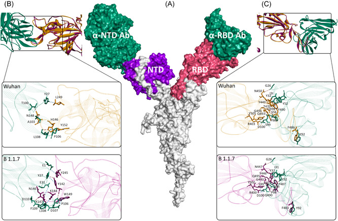
Figure 3.

Interaction between SARS‐CoV‐2 spike protein and antibodies. (A) Cartoon showing surface topology of antibodies (sea green) interaction with the NTD (purple) and RBD (pink) of the SARS‐CoV‐2 spike protein. Intermolecular interactions between, (B) NTD and, (C) RBD of SARS‐CoV‐2/Wuhan (gold) and SARS‐CoV‐2/B.1.1.7 (magenta) spike proteins in ribbon conformation. The enlarged views in each showing stick models of intermolecular interactions between the antibody residues (sea green) and amino acids of spike proteins of SARS‐CoV‐2/Wuhan (gold) and SARS‐CoV‐2/B.1.1.7 (magenta). Intermolecular hydrogen bonds and non‐hydrogen bond intermolecular interactions (electrostatic and hydrophobic) are shown with brown and blue‐dotted lines, respectively
