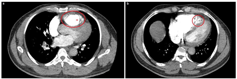
Figure 2.
Computed tomography scan, pulmonary embolism protocol. (a) The red circle indicates right ventricular dilatation, whereas the red arrow indicates the trabecular meshwork at the right ventricular apex. (b) The red circle indicates a more focused image of the right ventricular enlargement with marked sinusoids amidst the trabecular mesh.

