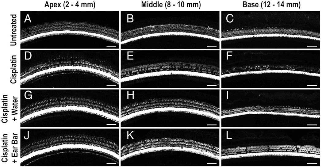
FIG. 3.

Hair cell confocal images. Representative confocal images are shown for apical (A, D, G, J) middle (B, E, H, K), and basal regions (C, F, I, L) of guinea pig cochleae for untreated, no-cisplatin control (A–C), cisplatin-only control (D–F), and cisplatin plus cooling (G–L). Less hair cell loss was observed for the animals undergoing ear cooling.
